 Movie
Movie Controller

[English] 日本語
 Yorodumi
- PDB-2d2r: Crystal structure of Helicobacter pylori Undecaprenyl Pyrophospha... -
+ Open data

- Basic information
| Entry | Database: PDB / ID: 2d2r | ||||||
|---|---|---|---|---|---|---|---|
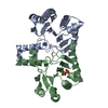
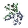
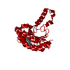
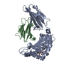
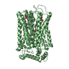
| Title | Crystal structure of Helicobacter pylori Undecaprenyl Pyrophosphate Synthase | ||||||
 Components Components | Undecaprenyl Pyrophosphate Synthase | ||||||
 Keywords Keywords | TRANSFERASE / prenyl / prenyltransferase / undecaprenyl | ||||||
| Function / homology |  Function and homology information Function and homology informationditrans,polycis-undecaprenyl-diphosphate synthase [(2E,6E)-farnesyl-diphosphate specific] activity / polyprenol biosynthetic process / Transferases; Transferring alkyl or aryl groups, other than methyl groups / magnesium ion binding / cytosol Similarity search - Function | ||||||
| Biological species |   Helicobacter pylori (bacteria) Helicobacter pylori (bacteria) | ||||||
| Method |  X-RAY DIFFRACTION / SYNCHROTRON / MOLECULAR REPLACEMENT / Resolution: 1.88 Å X-RAY DIFFRACTION / SYNCHROTRON / MOLECULAR REPLACEMENT / Resolution: 1.88 Å | ||||||
 Authors Authors | Kuo, C.J. / Guo, R.T. / Chen, C.L. / Ko, T.P. / Cheng, Y.S. / Cheng, Y.L. / Liang, P.H. / Wang, A.H.-J. | ||||||
 Citation Citation | Journal: J.Biomed.Biotechnol. / Year: 2008 Title: Structure-based inhibitors exhibit differential activities against Helicobacter pylori and Escherichia coli undecaprenyl pyrophosphate synthases. Authors: Kuo, C.J. / Guo, R.T. / Lu, I.L. / Liu, H.G. / Wu, S.Y. / Ko, T.P. / Wang, A.H. / Liang, P.H. | ||||||
| History |
|
- Structure visualization
| Structure viewer | Molecule: MolmilJmol/JSmol |
|---|
- Downloads & links
-Download
| PDBx/mmCIF format | 2d2r.cif.gz | 111.9 KB | Display | PDBx/mmCIF format |
|---|---|---|---|---|
| PDB format | pdb2d2r.ent.gz | 85.5 KB | Display | PDB format |
| PDBx/mmJSON format | 2d2r.json.gz | Tree view | PDBx/mmJSON format | |
| Others |  Other downloads Other downloads |
-Validation report
| Summary document | 2d2r_validation.pdf.gz | 411.1 KB | Display | wwPDB validaton report |
|---|---|---|---|---|
| Full document | 2d2r_full_validation.pdf.gz | 422.1 KB | Display | |
| Data in XML | 2d2r_validation.xml.gz | 12.2 KB | Display | |
| Data in CIF | 2d2r_validation.cif.gz | 20.5 KB | Display | |
| Arichive directory | https://data.pdbj.org/pub/pdb/validation_reports/d2/2d2rftp://data.pdbj.org/pub/pdb/validation_reports/d2/2d2r | HTTPS FTP |
-Related structure data
| Related structure data |  1x06S S: Starting model for refinement |
|---|---|
| Similar structure data |


-Links
 PDBj
PDBj- Assembly
| Deposited unit | 
| ||||||||
|---|---|---|---|---|---|---|---|---|---|
| 1 |
| ||||||||
| Unit cell |
|
-Components
| #1: Protein | Mass: 28575.924 Da / Num. of mol.: 2 / Mutation: C234A Source method: isolated from a genetically manipulated source Source: (gene. exp.) Helicobacter pylori (bacteria) / Strain: ATCC43504 / Plasmid: pET32 Xa/LIC / Species (production host): Escherichia coli / Production host: References: UniProt: P55984, ditrans,polycis-undecaprenyl-diphosphate synthase [(2E,6E)-farnesyl-diphosphate specific] #2: Water | ChemComp-HOH / |  Mass: 18.015 Da / Num. of mol.: 581 / Source method: isolated from a natural source / Formula: H2O Mass: 18.015 Da / Num. of mol.: 581 / Source method: isolated from a natural source / Formula: H2O |
|---|
-Experimental details
-Experiment
| Experiment | Method: X-RAY DIFFRACTION / Number of used crystals: 1 |
|---|
- Sample preparation
| Crystal | Density Matthews: 1.965 Å3/Da / Density % sol: 35 % |
|---|---|
| Crystal grow | Temperature: 298 K / Method: vapor diffusion, hanging drop / pH: 8 Details: KSCN, PEG600, PEG5KMME, pH 8.0, VAPOR DIFFUSION, HANGING DROP, temperature 298.0K |
-Data collection
| Diffraction | Mean temperature: 100 K |
|---|---|
| Diffraction source | Source: SYNCHROTRON / Site: NSRRC  / Beamline: BL17B2 / Wavelength: 1 Å / Beamline: BL17B2 / Wavelength: 1 Å |
| Detector | Type: RIGAKU RAXIS IV / Detector: IMAGE PLATE / Date: Feb 11, 2004 / Details: mirrors |
| Radiation | Monochromator: GRAPHITE / Protocol: SINGLE WAVELENGTH / Monochromatic (M) / Laue (L): M / Scattering type: x-ray |
| Radiation wavelength | Wavelength: 1 Å / Relative weight: 1 |
| Reflection | Resolution: 1.88→25 Å / Num. all: 37471 / Num. obs: 35917 / % possible obs: 95.59 % / Observed criterion σ(F): 0 / Observed criterion σ(I): 0 / Redundancy: 5.6 % / Rmerge(I) obs: 0.055 / Net I/σ(I): 30.7 |
| Reflection shell | Resolution: 1.88→1.95 Å / Redundancy: 5.6 % / Rmerge(I) obs: 0.433 / Mean I/σ(I) obs: 4.1 / Num. unique all: 3338 / % possible all: 90.3 |
- Processing
| Software |
| |||||||||||||||||||||||||
|---|---|---|---|---|---|---|---|---|---|---|---|---|---|---|---|---|---|---|---|---|---|---|---|---|---|---|
| Refinement | Method to determine structure: MOLECULAR REPLACEMENT Starting model: PDB ENTRY 1X06 Resolution: 1.88→25 Å / Cross valid method: THROUGHOUT / σ(F): 0 / σ(I): 0 / Stereochemistry target values: Engh & Huber
| |||||||||||||||||||||||||
| Refine analyze | Luzzati coordinate error obs: 0.21 Å / Luzzati d res low obs: 5 Å / Luzzati sigma a obs: 0.14 Å | |||||||||||||||||||||||||
| Refinement step | Cycle: LAST / Resolution: 1.88→25 Å
| |||||||||||||||||||||||||
| Refine LS restraints |
| |||||||||||||||||||||||||
| LS refinement shell | Resolution: 1.88→1.95 Å
|
