 Movie
Movie Controller

[English] 日本語
 Yorodumi
- PDB-3th8: Structure of E. coli undecaprenyl diphosphate synthase complexed ... -
+ Open data

- Basic information
| Entry | Database: PDB / ID: 3th8 | ||||||
|---|---|---|---|---|---|---|---|
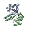
| Title | Structure of E. coli undecaprenyl diphosphate synthase complexed with BPH-1063 | ||||||
 Components Components | Undecaprenyl pyrophosphate synthase | ||||||
 Keywords Keywords | TRANSFERASE/TRANSFERASE INHIBITOR / all helices / prenyl transferase / farneyl diphosphate binding / Isopentenyl diphosphate binding / prenylation / TRANSFERASE-TRANSFERASE INHIBITOR complex | ||||||
| Function / homology |  Function and homology information Function and homology informationGram-negative-bacterium-type cell wall biogenesis / ditrans,polycis-undecaprenyl-diphosphate synthase [(2E,6E)-farnesyl-diphosphate specific] / ditrans,polycis-undecaprenyl-diphosphate synthase [(2E,6E)-farnesyl-diphosphate specific] activity / polyprenol biosynthetic process / small molecule binding / peptidoglycan biosynthetic process / cell wall organization / regulation of cell shape / cell division / magnesium ion binding ...Gram-negative-bacterium-type cell wall biogenesis / ditrans,polycis-undecaprenyl-diphosphate synthase [(2E,6E)-farnesyl-diphosphate specific] / ditrans,polycis-undecaprenyl-diphosphate synthase [(2E,6E)-farnesyl-diphosphate specific] activity / polyprenol biosynthetic process / small molecule binding / peptidoglycan biosynthetic process / cell wall organization / regulation of cell shape / cell division / magnesium ion binding / protein homodimerization activity / cytoplasm / cytosol Similarity search - Function | ||||||
| Biological species |  | ||||||
| Method |  X-RAY DIFFRACTION / SYNCHROTRON / MOLECULAR REPLACEMENT / Resolution: 2.114 Å X-RAY DIFFRACTION / SYNCHROTRON / MOLECULAR REPLACEMENT / Resolution: 2.114 Å | ||||||
 Authors Authors | Cao, R. / Zhu, W. / Zhang, Y. / Oldfield, E. | ||||||
 Citation Citation | Journal: ACS Med Chem Lett / Year: 2012 Title: HIV-1 Integrase Inhibitor-Inspired Antibacterials Targeting Isoprenoid Biosynthesis. Authors: Zhang, Y. / Lin, F.-Y. / Li, K. / Zhu, W. / Liu, Y.L. / Cao, R. / Pang, R. / Lee, E. / Axelson, J. / Hensler, M. / Wang, K. / Molohon, K.J. / Wang, Y. / Mitchell, D.A. / Nizet, V. / Oldfield, E. | ||||||
| History |
|
- Structure visualization
| Structure viewer | Molecule: MolmilJmol/JSmol |
|---|
- Downloads & links
-Download
| PDBx/mmCIF format | 3th8.cif.gz | 96.5 KB | Display | PDBx/mmCIF format |
|---|---|---|---|---|
| PDB format | pdb3th8.ent.gz | 73.4 KB | Display | PDB format |
| PDBx/mmJSON format | 3th8.json.gz | Tree view | PDBx/mmJSON format | |
| Others |  Other downloads Other downloads |
-Validation report
| Arichive directory | https://data.pdbj.org/pub/pdb/validation_reports/th/3th8ftp://data.pdbj.org/pub/pdb/validation_reports/th/3th8 | HTTPS FTP |
|---|
-Related structure data
| Related structure data | 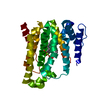 4f6vC 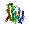 4f6xC 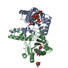 2e98S S: Starting model for refinement C: citing same article ( |
|---|---|
| Similar structure data |


-Links
 PDBj
PDBj- Assembly
| Deposited unit | 
| ||||||||
|---|---|---|---|---|---|---|---|---|---|
| 1 |
| ||||||||
| Unit cell |
|
-Components
| #1: Protein | Mass: 28481.127 Da / Num. of mol.: 2 Source method: isolated from a genetically manipulated source Source: (gene. exp.) References: UniProt: P60472, ditrans,polycis-undecaprenyl-diphosphate synthase [(2E,6E)-farnesyl-diphosphate specific] #2: Chemical | ChemComp-TH9 / ( |   Mass: 349.421 Da / Num. of mol.: 1 / Source method: obtained synthetically / Formula: C19H27NO5 Mass: 349.421 Da / Num. of mol.: 1 / Source method: obtained synthetically / Formula: C19H27NO5#3: Water | ChemComp-HOH / |  Mass: 18.015 Da / Num. of mol.: 63 / Source method: isolated from a natural source / Formula: H2O Mass: 18.015 Da / Num. of mol.: 63 / Source method: isolated from a natural source / Formula: H2O |
|---|
-Experimental details
-Experiment
| Experiment | Method: X-RAY DIFFRACTION / Number of used crystals: 1 |
|---|
- Sample preparation
| Crystal | Density Matthews: 1.85 Å3/Da / Density % sol: 33.64 % |
|---|---|
| Crystal grow | Temperature: 298.15 K / Method: vapor diffusion, hanging drop / pH: 7.5 Details: 5% PEG 4000, pH 7.5, VAPOR DIFFUSION, HANGING DROP, temperature 298.15K |
-Data collection
| Diffraction | Mean temperature: 123 K |
|---|---|
| Diffraction source | Source: SYNCHROTRON / Site: APS  / Beamline: 21-ID-F / Wavelength: 0.97872 Å / Beamline: 21-ID-F / Wavelength: 0.97872 Å |
| Detector | Type: MARMOSAIC 225 mm CCD / Detector: CCD / Date: Apr 3, 2010 |
| Radiation | Monochromator: C(111) / Protocol: SINGLE WAVELENGTH / Monochromatic (M) / Laue (L): M / Scattering type: x-ray |
| Radiation wavelength | Wavelength: 0.97872 Å / Relative weight: 1 |
| Reflection | Resolution: 2.114→50 Å / Num. obs: 27971 / % possible obs: 98.2 % / Observed criterion σ(F): 1 / Observed criterion σ(I): 1 |
| Reflection shell | Resolution: 2.114→2.16 Å / % possible all: 100 |
- Processing
| Software |
| |||||||||||||||||||||||||||||||||||||||||||||||||||||||||||||||||
|---|---|---|---|---|---|---|---|---|---|---|---|---|---|---|---|---|---|---|---|---|---|---|---|---|---|---|---|---|---|---|---|---|---|---|---|---|---|---|---|---|---|---|---|---|---|---|---|---|---|---|---|---|---|---|---|---|---|---|---|---|---|---|---|---|---|---|
| Refinement | Method to determine structure: MOLECULAR REPLACEMENT Starting model: PDB ENTRY 2E98 Resolution: 2.114→41.8 Å / Cor.coef. Fo:Fc: 0.911 / Cor.coef. Fo:Fc free: 0.889 / Occupancy max: 1 / Occupancy min: 1 / SU B: 6.312 / SU ML: 0.169 / Cross valid method: THROUGHOUT / σ(F): 0 / ESU R: 0.281 / ESU R Free: 0.241 / Stereochemistry target values: MAXIMUM LIKELIHOOD Details: HYDROGENS HAVE BEEN ADDED IN THE RIDING POSITIONS U VALUES : REFINED INDIVIDUALLY
| |||||||||||||||||||||||||||||||||||||||||||||||||||||||||||||||||
| Solvent computation | Ion probe radii: 0.8 Å / Shrinkage radii: 0.8 Å / VDW probe radii: 1.4 Å / Solvent model: MASK | |||||||||||||||||||||||||||||||||||||||||||||||||||||||||||||||||
| Displacement parameters | Biso max: 77.71 Å2 / Biso mean: 35.9978 Å2 / Biso min: 12.76 Å2
| |||||||||||||||||||||||||||||||||||||||||||||||||||||||||||||||||
| Refinement step | Cycle: LAST / Resolution: 2.114→41.8 Å
| |||||||||||||||||||||||||||||||||||||||||||||||||||||||||||||||||
| Refine LS restraints |
| |||||||||||||||||||||||||||||||||||||||||||||||||||||||||||||||||
| LS refinement shell | Resolution: 2.114→2.169 Å / Total num. of bins used: 20
|
