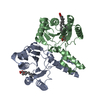
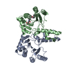
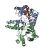
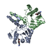
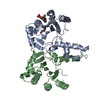
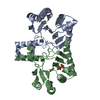
[English] 日本語
Yorodumi
- PDB-5zhe: STRUCTURE OF E. COLI UNDECAPRENYL DIPHOSPHATE SYNTHASE IN COMPLEX... -

+
Open data


ID or keywords:

Loading...

-
Basic information

Entry
Database: PDB / ID: 5zhe
TitleSTRUCTURE OF E. COLI UNDECAPRENYL DIPHOSPHATE SYNTHASE IN COMPLEX WITH BPH-981
ComponentsDitrans,polycis-undecaprenyl-diphosphate synthase ((2E,6E)-farnesyl-diphosphate specific)
KeywordsTRANSFERASE/INHIBITOR / metal-binding / substrate binding / acidocalcisomal pyrophosphatase / TRANSFERASE-INHIBITOR complex
Function / homology
Function and homology information


Gram-negative-bacterium-type cell wall biogenesis / ditrans,polycis-undecaprenyl-diphosphate synthase [(2E,6E)-farnesyl-diphosphate specific] / di-trans,poly-cis-undecaprenyl-diphosphate synthase activity / polyprenol biosynthetic process / small molecule binding / peptidoglycan biosynthetic process / cell wall organization / regulation of cell shape / cell division / magnesium ion binding ...Gram-negative-bacterium-type cell wall biogenesis / ditrans,polycis-undecaprenyl-diphosphate synthase [(2E,6E)-farnesyl-diphosphate specific] / di-trans,poly-cis-undecaprenyl-diphosphate synthase activity / polyprenol biosynthetic process / small molecule binding / peptidoglycan biosynthetic process / cell wall organization / regulation of cell shape / cell division / magnesium ion binding / protein homodimerization activity / cytoplasm / cytosol
Similarity search - Function
Undecaprenyl pyrophosphate synthetase / Decaprenyl diphosphate synthase-like / Di-trans-poly-cis-decaprenylcistransferase-like, conserved site / Undecaprenyl pyrophosphate synthase family signature. / Decaprenyl diphosphate synthase-like / Putative undecaprenyl diphosphate synthase / Decaprenyl diphosphate synthase-like superfamily / 3-Layer(aba) Sandwich / Alpha Beta
Similarity search - Domain/homology
2-hydroxy-6-(tetradecyloxy)benzoic acid / Ditrans,polycis-undecaprenyl-diphosphate synthase ((2E,6E)-farnesyl-diphosphate specific)
Similarity search - Component
Biological speciesEscherichia coli K-12 (bacteria)
MethodX-RAY DIFFRACTION / SYNCHROTRON / MOLECULAR REPLACEMENT / Resolution: 2.18 Å
AuthorsGao, J. / Liu, W.D. / Zheng, Y.Y. / Ko, T.P. / Chen, C.C. / Guo, R.T.
CitationJournal: J.Med.Chem. / Year: 2019
Title: Discovery of Lipophilic Bisphosphonates That Target Bacterial Cell Wall and Quinone Biosynthesis.
Authors: Malwal, S.R. / Chen, L. / Hicks, H. / Qu, F. / Liu, W. / Shillo, A. / Law, W.X. / Zhang, J. / Chandnani, N. / Han, X. / Zheng, Y. / Chen, C.C. / Guo, R.T. / AbdelKhalek, A. / Seleem, M.N. / Oldfield, E.
History
DepositionMar 13, 2018Deposition site: PDBJ / Processing site: PDBJ
Revision 1.0Mar 13, 2019Provider: repository / Type: Initial release
Revision 1.1Sep 25, 2019Group: Data collection / Database references / Category: citation / citation_author
Item: _citation.country / _citation.journal_abbrev ..._citation.country / _citation.journal_abbrev / _citation.journal_id_ASTM / _citation.journal_id_CSD / _citation.journal_id_ISSN / _citation.journal_volume / _citation.page_first / _citation.page_last / _citation.pdbx_database_id_DOI / _citation.pdbx_database_id_PubMed / _citation.title / _citation.year
Revision 1.2Nov 22, 2023Group: Data collection / Database references / Refinement description
Category: chem_comp_atom / chem_comp_bond ...chem_comp_atom / chem_comp_bond / database_2 / pdbx_initial_refinement_model
Item: _database_2.pdbx_DOI / _database_2.pdbx_database_accession

-
Structure visualization

Structure viewerMolecule:
MolmilJmol/JSmol

Downloads & links

-
Assembly

Deposited unit
A: Ditrans,polycis-undecaprenyl-diphosphate synthase ((2E,6E)-farnesyl-diphosphate specific)
B: Ditrans,polycis-undecaprenyl-diphosphate synthase ((2E,6E)-farnesyl-diphosphate specific)
hetero molecules


Theoretical massNumber of molelcules
Total (without water)57,3133
Polymers56,9622
Non-polymers3501
Water2,342130
1


  • Idetical with deposited unit
  • defined by software
  • Evidence: gel filtration
TypeNameSymmetry operationNumber
identity operation1_555x,y,z1
Buried area3360 Å2
ΔGint-15 kcal/mol
Surface area19710 Å2
MethodPISA
Unit cell
Length a, b, c (Å)63.713, 68.449, 109.253
Angle α, β, γ (deg.)90.00, 90.00, 90.00
Int Tables number19
Space group name H-MP212121

-
Components

#1: Protein Ditrans,polycis-undecaprenyl-diphosphate synthase ((2E,6E)-farnesyl-diphosphate specific) / Ditrans / polycis-undecaprenylcistransferase


Mass: 28481.127 Da / Num. of mol.: 2
Source method: isolated from a genetically manipulated source
Source: (gene. exp.) Escherichia coli K-12 (bacteria) / Strain: K-12 / Gene: ispU, rth, uppS, yaeS, b0174, JW0169 / Plasmid: pET32EK/LIC / Production host: Escherichia coli BL21(DE3) (bacteria) / Strain (production host): BL21(DE3)
References: UniProt: P60472, ditrans,polycis-undecaprenyl-diphosphate synthase [(2E,6E)-farnesyl-diphosphate specific]
#2: Chemical ChemComp-HJX / 2-hydroxy-6-(tetradecyloxy)benzoic acid / BPH-981


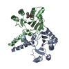
Mass: 350.492 Da / Num. of mol.: 1 / Source method: obtained synthetically / Formula: C21H34O4
#3: Water ChemComp-HOH / water


Mass: 18.015 Da / Num. of mol.: 130 / Source method: isolated from a natural source / Formula: H2O

-
Experimental details

-
Experiment

ExperimentMethod: X-RAY DIFFRACTION / Number of used crystals: 1

-
Sample preparation

CrystalDensity Matthews: 2.09 Å3/Da / Density % sol: 41.18 % / Mosaicity: 0.41 °
Crystal growTemperature: 295 K / Method: vapor diffusion, sitting drop / pH: 7.5 / Details: 20 % ETHYLENE GLYCOL, 2-5% PEG 35K

-
Data collection

DiffractionMean temperature: 100 K
Diffraction sourceSource: SYNCHROTRON / Site: NSRRC / Beamline: BL13B1 / Wavelength: 1 Å
DetectorType: ADSC QUANTUM 315 / Detector: CCD / Date: Feb 15, 2013
RadiationMonochromator: GRAPHITE / Protocol: SINGLE WAVELENGTH / Monochromatic (M) / Laue (L): M / Scattering type: x-ray
Radiation wavelengthWavelength: 1 Å / Relative weight: 1
ReflectionResolution: 2.18→25 Å / Num. obs: 25466 / % possible obs: 99.5 % / Redundancy: 6.3 % / Rmerge(I) obs: 0.044 / Χ2: 0.827 / Net I/σ(I): 18.4 / Num. measured all: 160471
Reflection shell
Resolution (Å)Redundancy (%)Rmerge(I) obsNum. unique allΧ2Diffraction-ID% possible all
2.18-2.2650.2624350.548197.6
2.26-2.3560.24125090.557199.8
2.35-2.456.60.16625300.5741100
2.45-2.586.70.12825150.6111100
2.58-2.756.70.08825510.697199.9
2.75-2.966.70.06125120.815199.9
2.96-3.256.60.04425610.9731100
3.25-3.726.50.03725821.11100
3.72-4.696.30.04125960.9921100
4.69-2560.0326751.317197.6

-
Processing

Software
NameVersionClassification
REFMAC5.8.0189refinement
HKL-2000data scaling
PDB_EXTRACT3.22data extraction
HKL-2000data reduction
PHASERphasing
RefinementMethod to determine structure: MOLECULAR REPLACEMENT
Starting model: 3WYJ
Resolution: 2.18→25 Å / Cor.coef. Fo:Fc: 0.95 / Cor.coef. Fo:Fc free: 0.926 / SU B: 6.147 / SU ML: 0.158 / Cross valid method: THROUGHOUT / ESU R: 0.254 / ESU R Free: 0.218 / Details: HYDROGENS HAVE BEEN ADDED IN THE RIDING POSITIONS
RfactorNum. reflection% reflectionSelection details
Rfree0.26308 1166 4.6 %RANDOM
Rwork0.20309 ---
obs0.20586 24247 99.3 %-
Solvent computationIon probe radii: 0.8 Å / Shrinkage radii: 0.8 Å / VDW probe radii: 1.2 Å
Displacement parametersBiso mean: 52.869 Å2
Baniso -1Baniso -2Baniso -3
1--1.36 Å2-0 Å20 Å2
2--1.3 Å2-0 Å2
3---0.06 Å2
Refinement stepCycle: 1 / Resolution: 2.18→25 Å
ProteinNucleic acidLigandSolventTotal
Num. atoms3223 0 25 130 3378
Refine LS restraints
Refine-IDTypeDev idealDev ideal targetNumber
X-RAY DIFFRACTIONr_bond_refined_d0.0120.0193328
X-RAY DIFFRACTIONr_bond_other_d0.0010.023069
X-RAY DIFFRACTIONr_angle_refined_deg1.7281.9254483
X-RAY DIFFRACTIONr_angle_other_deg0.92537062
X-RAY DIFFRACTIONr_dihedral_angle_1_deg6.6535403
X-RAY DIFFRACTIONr_dihedral_angle_2_deg36.5323.39177
X-RAY DIFFRACTIONr_dihedral_angle_3_deg19.3615558
X-RAY DIFFRACTIONr_dihedral_angle_4_deg24.8921533
X-RAY DIFFRACTIONr_chiral_restr0.1050.2477
X-RAY DIFFRACTIONr_gen_planes_refined0.0110.023754
X-RAY DIFFRACTIONr_gen_planes_other0.0020.02743
X-RAY DIFFRACTIONr_nbd_refined
X-RAY DIFFRACTIONr_nbd_other
X-RAY DIFFRACTIONr_nbtor_refined
X-RAY DIFFRACTIONr_nbtor_other
X-RAY DIFFRACTIONr_xyhbond_nbd_refined
X-RAY DIFFRACTIONr_xyhbond_nbd_other
X-RAY DIFFRACTIONr_metal_ion_refined
X-RAY DIFFRACTIONr_metal_ion_other
X-RAY DIFFRACTIONr_symmetry_vdw_refined
X-RAY DIFFRACTIONr_symmetry_vdw_other
X-RAY DIFFRACTIONr_symmetry_hbond_refined
X-RAY DIFFRACTIONr_symmetry_hbond_other
X-RAY DIFFRACTIONr_symmetry_metal_ion_refined
X-RAY DIFFRACTIONr_symmetry_metal_ion_other
X-RAY DIFFRACTIONr_mcbond_it6.0474.8791627
X-RAY DIFFRACTIONr_mcbond_other6.0484.8781626
X-RAY DIFFRACTIONr_mcangle_it7.8937.2722025
X-RAY DIFFRACTIONr_mcangle_other7.8917.2732026
X-RAY DIFFRACTIONr_scbond_it7.4215.7851701
X-RAY DIFFRACTIONr_scbond_other7.3935.7741698
X-RAY DIFFRACTIONr_scangle_it
X-RAY DIFFRACTIONr_scangle_other10.5068.3352459
X-RAY DIFFRACTIONr_long_range_B_refined13.5692.88213717
X-RAY DIFFRACTIONr_long_range_B_other13.55992.85713673
X-RAY DIFFRACTIONr_rigid_bond_restr
X-RAY DIFFRACTIONr_sphericity_free
X-RAY DIFFRACTIONr_sphericity_bonded
LS refinement shellResolution: 2.18→2.236 Å / Total num. of bins used: 20
RfactorNum. reflection% reflection
Rfree0.342 86 -
Rwork0.237 1660 -
obs--96.15 %

+
About Yorodumi

-
News

-
Feb 9, 2022. New format data for meta-information of EMDB entries

New format data for meta-information of EMDB entries

  • Version 3 of the EMDB header file is now the official format.
  • The previous official version 1.9 will be removed from the archive.

Related info.:EMDB header

External links:wwPDB to switch to version 3 of the EMDB data model

-
Aug 12, 2020. Covid-19 info

Covid-19 info

URL: https://pdbjlc1.pdbj.org/emnavi/covid19.php

New page: Covid-19 featured information page in EM Navigator.

Related info.:Covid-19 info / Mar 5, 2020. Novel coronavirus structure data

+
Mar 5, 2020. Novel coronavirus structure data

Novel coronavirus structure data

Related info.:Yorodumi Speices / Aug 12, 2020. Covid-19 info

External links:COVID-19 featured content - PDBj / Molecule of the Month (242):Coronavirus Proteases

+
Jan 31, 2019. EMDB accession codes are about to change! (news from PDBe EMDB page)

EMDB accession codes are about to change! (news from PDBe EMDB page)

  • The allocation of 4 digits for EMDB accession codes will soon come to an end. Whilst these codes will remain in use, new EMDB accession codes will include an additional digit and will expand incrementally as the available range of codes is exhausted. The current 4-digit format prefixed with “EMD-” (i.e. EMD-XXXX) will advance to a 5-digit format (i.e. EMD-XXXXX), and so on. It is currently estimated that the 4-digit codes will be depleted around Spring 2019, at which point the 5-digit format will come into force.
  • The EM Navigator/Yorodumi systems omit the EMD- prefix.

Related info.:Q: What is EMD? / ID/Accession-code notation in Yorodumi/EM Navigator

External links:EMDB Accession Codes are Changing Soon! / Contact to PDBj

+
Jul 12, 2017. Major update of PDB

Major update of PDB

  • wwPDB released updated PDB data conforming to the new PDBx/mmCIF dictionary.
  • This is a major update changing the version number from 4 to 5, and with Remediation, in which all the entries are updated.
  • In this update, many items about electron microscopy experimental information are reorganized (e.g. em_software).
  • Now, EM Navigator and Yorodumi are based on the updated data.

External links:wwPDB Remediation / Enriched Model Files Conforming to OneDep Data Standards Now Available in the PDB FTP Archive

-
Yorodumi

Thousand views of thousand structures

  • Yorodumi is a browser for structure data from EMDB, PDB, SASBDB, etc.
  • This page is also the successor to EM Navigator detail page, and also detail information page/front-end page for Omokage search.
  • The word "yorodu" (or yorozu) is an old Japanese word meaning "ten thousand". "mi" (miru) is to see.

Related info.:EMDB / PDB / SASBDB / Comparison of 3 databanks / Yorodumi Search / Aug 31, 2016. New EM Navigator & Yorodumi / Yorodumi Papers / Jmol/JSmol / Function and homology information / Changes in new EM Navigator and Yorodumi

Read more