[English] 日本語
Yorodumi
- PDB-2cx4: Crystal structure of a bacterioferritin comigratory protein perox... -

+
Open data


ID or keywords:

Loading...

-
Basic information

Entry
Database: PDB / ID: 2cx4
TitleCrystal structure of a bacterioferritin comigratory protein peroxiredoxin from the Aeropyrum pernix K1 (form-2 crystal)
Componentsbacterioferritin comigratory protein
KeywordsOXIDOREDUCTASE / antioxidant enzyme / reactive oxygen species / thioredoxin fold / structural genomics / NPPSFA / National Project on Protein Structural and Functional Analyses / RIKEN Structural Genomics/Proteomics Initiative / RSGI
Function / homology
Function and homology information


peroxidase activity
Similarity search - Function
Peroxiredoxin, AhpC-type / Alkyl hydroperoxide reductase subunit C/ Thiol specific antioxidant / AhpC/TSA family / Thioredoxin domain profile. / Thioredoxin domain / Glutaredoxin / Glutaredoxin / Thioredoxin-like superfamily / 3-Layer(aba) Sandwich / Alpha Beta
Similarity search - Domain/homology
: / Thiol peroxidase
Similarity search - Component
Biological speciesAeropyrum pernix (archaea)
MethodX-RAY DIFFRACTION / SYNCHROTRON / MAD / Resolution: 2.3 Å
AuthorsMizohata, E. / Murayama, K. / Idaka, M. / Tatsuguchi, A. / Terada, T. / Shirouzu, M. / Yokoyama, S. / RIKEN Structural Genomics/Proteomics Initiative (RSGI)
CitationJournal: To be Published
Title: Crystal structure of a bacterioferritin comigratory protein peroxiredoxin from the Aeropyrum pernix K1 (form-2 crystal)
Authors: Mizohata, E. / Murayama, K. / Idaka, M. / Tatsuguchi, A. / Terada, T. / Shirouzu, M. / Yokoyama, S.
History
DepositionJun 27, 2005Deposition site: PDBJ / Processing site: PDBJ
Revision 1.0Dec 27, 2005Provider: repository / Type: Initial release
Revision 1.1Apr 30, 2008Group: Version format compliance
Revision 1.2Jul 13, 2011Group: Source and taxonomy / Version format compliance

-
Structure visualization

Structure viewerMolecule:
MolmilJmol/JSmol

Downloads & links

-
Assembly

Deposited unit
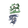
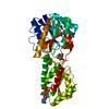
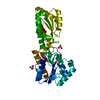
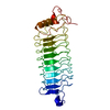
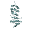
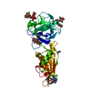
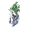
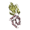
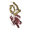
A: bacterioferritin comigratory protein
B: bacterioferritin comigratory protein
C: bacterioferritin comigratory protein
D: bacterioferritin comigratory protein
E: bacterioferritin comigratory protein
F: bacterioferritin comigratory protein
G: bacterioferritin comigratory protein
H: bacterioferritin comigratory protein


Theoretical massNumber of molelcules
Total (without water)150,6828
Polymers150,6828
Non-polymers00
Water7,332407
1
A: bacterioferritin comigratory protein
B: bacterioferritin comigratory protein


Theoretical massNumber of molelcules
Total (without water)37,6712
Polymers37,6712
Non-polymers00
Water362
TypeNameSymmetry operationNumber
identity operation1_555x,y,z1
Buried area2000 Å2
ΔGint-14 kcal/mol
Surface area14320 Å2
MethodPISA
2
C: bacterioferritin comigratory protein
D: bacterioferritin comigratory protein


Theoretical massNumber of molelcules
Total (without water)37,6712
Polymers37,6712
Non-polymers00
Water362
TypeNameSymmetry operationNumber
identity operation1_555x,y,z1
Buried area1970 Å2
ΔGint-16 kcal/mol
Surface area14270 Å2
MethodPISA
3
E: bacterioferritin comigratory protein
F: bacterioferritin comigratory protein


Theoretical massNumber of molelcules
Total (without water)37,6712
Polymers37,6712
Non-polymers00
Water362
TypeNameSymmetry operationNumber
identity operation1_555x,y,z1
Buried area2080 Å2
ΔGint-14 kcal/mol
Surface area14430 Å2
MethodPISA
4
G: bacterioferritin comigratory protein
H: bacterioferritin comigratory protein


Theoretical massNumber of molelcules
Total (without water)37,6712
Polymers37,6712
Non-polymers00
Water362
TypeNameSymmetry operationNumber
identity operation1_555x,y,z1
Buried area2010 Å2
ΔGint-13 kcal/mol
Surface area14300 Å2
MethodPISA
Unit cell
Length a, b, c (Å)132.479, 132.479, 106.588
Angle α, β, γ (deg.)90.00, 90.00, 90.00
Int Tables number76
Space group name H-MP41

-
Components

#1: Protein
bacterioferritin comigratory protein / bacterioferritin comigratory protein peroxiredoxin


Mass: 18835.289 Da / Num. of mol.: 8
Source method: isolated from a genetically manipulated source
Source: (gene. exp.) Aeropyrum pernix (archaea) / Strain: K1 / Gene: APE2125 / Plasmid: pET11a / Production host: Escherichia coli (E. coli) / Strain (production host): B834(DE3)
References: GenBank: 14601861, UniProt: Q9YA14*PLUS, peroxiredoxin
#2: Water ChemComp-HOH / water


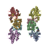
Mass: 18.015 Da / Num. of mol.: 407 / Source method: isolated from a natural source / Formula: H2O

-
Experimental details

-
Experiment

ExperimentMethod: X-RAY DIFFRACTION / Number of used crystals: 1

-
Sample preparation

CrystalDensity Matthews: 3.13 Å3/Da / Density % sol: 60 %
Crystal growTemperature: 285 K / Method: vapor diffusion, sitting drop / pH: 7
Details: ammonium sulfate, MES, Tris, sodium chloride, dithiothreitol, pH 7, VAPOR DIFFUSION, SITTING DROP, temperature 285K

-
Data collection

DiffractionMean temperature: 100 K
Diffraction sourceSource: SYNCHROTRON / Site: SPring-8 / Beamline: BL26B1 / Wavelength: 0.96400, 0.97884, 0.97946
DetectorType: RIGAKU JUPITER 210 / Detector: CCD / Date: Apr 6, 2004
RadiationProtocol: MAD / Monochromatic (M) / Laue (L): M / Scattering type: x-ray
Radiation wavelength
IDWavelength (Å)Relative weight
10.9641
20.978841
30.979461
ReflectionResolution: 2.3→50 Å / Num. obs: 143980 / Biso Wilson estimate: 20 Å2

-
Processing

Software
NameVersionClassification
CNS1.1refinement
HKL-2000data reduction
SCALEPACKdata scaling
SOLVEphasing
RefinementMethod to determine structure: MAD / Resolution: 2.3→46.32 Å / Isotropic thermal model: RESTRAINED / Cross valid method: THROUGHOUT / σ(F): 0
Details: The crystal form is twinned by the operator k, h, -l, twinning fraction 0.5
RfactorNum. reflection% reflectionSelection details
Rfree0.263 --RANDOM
Rwork0.203 ---
obs0.203 143980 89.6 %-
Displacement parametersBiso mean: 49.1 Å2
Refine analyze
FreeObs
Luzzati coordinate error0.38 Å0.31 Å
Luzzati d res low-5 Å
Luzzati sigma a0.44 Å0.48 Å
Refinement stepCycle: LAST / Resolution: 2.3→46.32 Å
ProteinNucleic acidLigandSolventTotal
Num. atoms10328 0 0 407 10735
Refine LS restraints
Refine-IDTypeDev ideal
X-RAY DIFFRACTIONc_bond_d0.007
X-RAY DIFFRACTIONc_bond_d_na
X-RAY DIFFRACTIONc_bond_d_prot
X-RAY DIFFRACTIONc_angle_d
X-RAY DIFFRACTIONc_angle_d_na
X-RAY DIFFRACTIONc_angle_d_prot
X-RAY DIFFRACTIONc_angle_deg1.1
X-RAY DIFFRACTIONc_angle_deg_na
X-RAY DIFFRACTIONc_angle_deg_prot
X-RAY DIFFRACTIONc_dihedral_angle_d23.6
X-RAY DIFFRACTIONc_dihedral_angle_d_na
X-RAY DIFFRACTIONc_dihedral_angle_d_prot
X-RAY DIFFRACTIONc_improper_angle_d0.79
X-RAY DIFFRACTIONc_improper_angle_d_na
X-RAY DIFFRACTIONc_improper_angle_d_prot
X-RAY DIFFRACTIONc_mcbond_it
X-RAY DIFFRACTIONc_mcangle_it
X-RAY DIFFRACTIONc_scbond_it
X-RAY DIFFRACTIONc_scangle_it
LS refinement shellResolution: 2.3→2.38 Å / Total num. of bins used: 10
RfactorNum. reflection% reflection
Rfree0.319 --
Rwork0.322 8442 -
obs--54.8 %

+
About Yorodumi

-
News

-
Feb 9, 2022. New format data for meta-information of EMDB entries

New format data for meta-information of EMDB entries

  • Version 3 of the EMDB header file is now the official format.
  • The previous official version 1.9 will be removed from the archive.

Related info.:EMDB header

External links:wwPDB to switch to version 3 of the EMDB data model

-
Aug 12, 2020. Covid-19 info

Covid-19 info

URL: https://pdbjlvh1.pdbj.org/emnavi/covid19.php

New page: Covid-19 featured information page in EM Navigator.

Related info.:Covid-19 info / Mar 5, 2020. Novel coronavirus structure data

+
Mar 5, 2020. Novel coronavirus structure data

Novel coronavirus structure data

Related info.:Yorodumi Speices / Aug 12, 2020. Covid-19 info

External links:COVID-19 featured content - PDBj / Molecule of the Month (242):Coronavirus Proteases

+
Jan 31, 2019. EMDB accession codes are about to change! (news from PDBe EMDB page)

EMDB accession codes are about to change! (news from PDBe EMDB page)

  • The allocation of 4 digits for EMDB accession codes will soon come to an end. Whilst these codes will remain in use, new EMDB accession codes will include an additional digit and will expand incrementally as the available range of codes is exhausted. The current 4-digit format prefixed with “EMD-” (i.e. EMD-XXXX) will advance to a 5-digit format (i.e. EMD-XXXXX), and so on. It is currently estimated that the 4-digit codes will be depleted around Spring 2019, at which point the 5-digit format will come into force.
  • The EM Navigator/Yorodumi systems omit the EMD- prefix.

Related info.:Q: What is EMD? / ID/Accession-code notation in Yorodumi/EM Navigator

External links:EMDB Accession Codes are Changing Soon! / Contact to PDBj

+
Jul 12, 2017. Major update of PDB

Major update of PDB

  • wwPDB released updated PDB data conforming to the new PDBx/mmCIF dictionary.
  • This is a major update changing the version number from 4 to 5, and with Remediation, in which all the entries are updated.
  • In this update, many items about electron microscopy experimental information are reorganized (e.g. em_software).
  • Now, EM Navigator and Yorodumi are based on the updated data.

External links:wwPDB Remediation / Enriched Model Files Conforming to OneDep Data Standards Now Available in the PDB FTP Archive

-
Yorodumi

Thousand views of thousand structures

  • Yorodumi is a browser for structure data from EMDB, PDB, SASBDB, etc.
  • This page is also the successor to EM Navigator detail page, and also detail information page/front-end page for Omokage search.
  • The word "yorodu" (or yorozu) is an old Japanese word meaning "ten thousand". "mi" (miru) is to see.

Related info.:EMDB / PDB / SASBDB / Comparison of 3 databanks / Yorodumi Search / Aug 31, 2016. New EM Navigator & Yorodumi / Yorodumi Papers / Jmol/JSmol / Function and homology information / Changes in new EM Navigator and Yorodumi

Read more