 Movie
Movie Controller


+ Open data

- Basic information
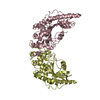
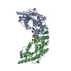
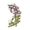
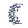
| Entry | Database: PDB / ID: 2crk | ||||||
|---|---|---|---|---|---|---|---|
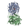
| Title | MUSCLE CREATINE KINASE | ||||||
 Components Components | PROTEIN (CREATINE KINASE) | ||||||
 Keywords Keywords | TRANSFERASE / CREATINE KINASE | ||||||
| Function / homology |  Function and homology information Function and homology informationcreatine kinase / phosphocreatine biosynthetic process / creatine kinase activity / response to heat / : / ATP binding Similarity search - Function | ||||||
| Biological species |  | ||||||
| Method |  X-RAY DIFFRACTION / MIR / Resolution: 2.35 Å X-RAY DIFFRACTION / MIR / Resolution: 2.35 Å | ||||||
 Authors Authors | Rao, J.K. / Bujacz, G. / Wlodawer, A. | ||||||
 Citation Citation | Journal: FEBS Lett. / Year: 1998 Title: Crystal structure of rabbit muscle creatine kinase. Authors: Rao, J.K. / Bujacz, G. / Wlodawer, A. | ||||||
| History |
|
- Structure visualization
| Structure viewer | Molecule: MolmilJmol/JSmol |
|---|
- Downloads & links
-Download
| PDBx/mmCIF format | 2crk.cif.gz | 95.3 KB | Display | PDBx/mmCIF format |
|---|---|---|---|---|
| PDB format | pdb2crk.ent.gz | 72.5 KB | Display | PDB format |
| PDBx/mmJSON format | 2crk.json.gz | Tree view | PDBx/mmJSON format | |
| Others |  Other downloads Other downloads |
-Validation report
| Arichive directory | https://data.pdbj.org/pub/pdb/validation_reports/cr/2crkftp://data.pdbj.org/pub/pdb/validation_reports/cr/2crk | HTTPS FTP |
|---|
-Related structure data
| Related structure data | 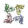 1crkS S: Starting model for refinement |
|---|---|
| Similar structure data |


-Links
 PDBj
PDBj
- Assembly
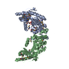
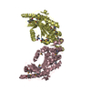
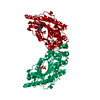
| Deposited unit | 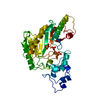
| ||||||||
|---|---|---|---|---|---|---|---|---|---|
| 1 | 
| ||||||||
| Unit cell |
|
-Components
| #1: Protein | Mass: 43301.227 Da / Num. of mol.: 1 / Source method: isolated from a natural source / Source: (natural) | ||
|---|---|---|---|
| #2: Chemical | ChemComp-SO4 /   Mass: 96.063 Da / Num. of mol.: 4 / Source method: obtained synthetically / Formula: SO4 Mass: 96.063 Da / Num. of mol.: 4 / Source method: obtained synthetically / Formula: SO4#3: Water | ChemComp-HOH / |  Mass: 18.015 Da / Num. of mol.: 431 / Source method: isolated from a natural source / Formula: H2O Mass: 18.015 Da / Num. of mol.: 431 / Source method: isolated from a natural source / Formula: H2O |
-Experimental details
-Experiment
| Experiment | Method: X-RAY DIFFRACTION |
|---|
- Sample preparation
| Crystal | Density Matthews: 4.2 Å3/Da / Density % sol: 68 % | |||||||||||||||||||||||||
|---|---|---|---|---|---|---|---|---|---|---|---|---|---|---|---|---|---|---|---|---|---|---|---|---|---|---|
| Crystal grow | pH: 7.2 Details: 55% TO 60% SATURATED AMMONIUM SULFATE, 2.5% PEG400 IN HEPES BUFFER AT PH 7.2, ABOUT 5MM/MG PROTEIN CONCENTRATION | |||||||||||||||||||||||||
| Crystal | *PLUS | |||||||||||||||||||||||||
| Crystal grow | *PLUS Method: standing drop | |||||||||||||||||||||||||
| Components of the solutions | *PLUS
|
-Data collection
| Diffraction | Mean temperature: 120 K |
|---|---|
| Diffraction source | Source: ROTATING ANODE / Type: ENRAF-NONIUS FR591 / Wavelength: 1.5418 |
| Detector | Type: MAC Science DIP-2020 / Detector: IMAGE PLATE / Date: Oct 15, 1996 / Details: FRANKS DOUBLE MIRROR OPTICS |
| Radiation | Monochromator: PT AND NI COATED GLASS MIRRORS / Protocol: SINGLE WAVELENGTH / Monochromatic (M) / Laue (L): M / Scattering type: x-ray |
| Radiation wavelength | Wavelength: 1.5418 Å / Relative weight: 1 |
| Reflection | Resolution: 2.35→20 Å / Num. obs: 29042 / % possible obs: 96.6 % / Observed criterion σ(I): 1 / Redundancy: 9.4 % / Rsym value: 0.101 / Net I/σ(I): 16.45 |
| Reflection shell | Resolution: 2.35→2.39 Å / Redundancy: 5.3 % / Mean I/σ(I) obs: 3.78 / Rsym value: 0.339 / % possible all: 89.2 |
| Reflection | *PLUS Rmerge(I) obs: 0.101 |
- Processing
| Software |
| ||||||||||||||||||||||||||||||||||||||||||||||||||||||||||||
|---|---|---|---|---|---|---|---|---|---|---|---|---|---|---|---|---|---|---|---|---|---|---|---|---|---|---|---|---|---|---|---|---|---|---|---|---|---|---|---|---|---|---|---|---|---|---|---|---|---|---|---|---|---|---|---|---|---|---|---|---|---|
| Refinement | Method to determine structure: MIR Starting model: 1CRK Resolution: 2.35→7 Å / Cross valid method: THROUGHOUT / σ(F): 2
| ||||||||||||||||||||||||||||||||||||||||||||||||||||||||||||
| Displacement parameters | Biso mean: 26.3 Å2 | ||||||||||||||||||||||||||||||||||||||||||||||||||||||||||||
| Refine analyze | Luzzati coordinate error obs: 0.25 Å | ||||||||||||||||||||||||||||||||||||||||||||||||||||||||||||
| Refinement step | Cycle: LAST / Resolution: 2.35→7 Å
| ||||||||||||||||||||||||||||||||||||||||||||||||||||||||||||
| Refine LS restraints |
| ||||||||||||||||||||||||||||||||||||||||||||||||||||||||||||
| LS refinement shell | Resolution: 2.35→2.43 Å / Total num. of bins used: 10 /
| ||||||||||||||||||||||||||||||||||||||||||||||||||||||||||||
| Xplor file | Serial no: 1 / Param file: PARAM19X.PRO / Topol file: TOPH19X.PRO | ||||||||||||||||||||||||||||||||||||||||||||||||||||||||||||
| Software | *PLUS Name: X-PLOR / Classification: refinement | ||||||||||||||||||||||||||||||||||||||||||||||||||||||||||||
| Refinement | *PLUS Lowest resolution: 7 Å / σ(F): 2 / Num. reflection Rfree: 2943 / % reflection Rfree: 9 % | ||||||||||||||||||||||||||||||||||||||||||||||||||||||||||||
| Solvent computation | *PLUS | ||||||||||||||||||||||||||||||||||||||||||||||||||||||||||||
| Displacement parameters | *PLUS Biso mean: 26.3 Å2 | ||||||||||||||||||||||||||||||||||||||||||||||||||||||||||||
| Refine LS restraints | *PLUS
| ||||||||||||||||||||||||||||||||||||||||||||||||||||||||||||
| LS refinement shell | *PLUS Rfactor Rfree: 0.285 / % reflection Rfree: 9 % / Rfactor Rwork: 0.377 |
