 Movie
Movie Controller


+ Open data

- Basic information
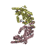
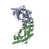
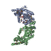
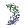
| Entry | Database: PDB / ID: 1i0e | ||||||
|---|---|---|---|---|---|---|---|
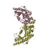
| Title | CRYSTAL STRUCTURE OF CREATINE KINASE FROM HUMAN MUSCLE | ||||||
 Components Components | CREATINE KINASE,M CHAIN | ||||||
 Keywords Keywords | TRANSFERASE / Double helix / dimer | ||||||
| Function / homology |  Function and homology information Function and homology informationcreatine kinase / phosphocreatine biosynthetic process / Creatine metabolism / creatine kinase activity / : / ATP binding / cytosol Similarity search - Function | ||||||
| Biological species |  Homo sapiens (human) Homo sapiens (human) | ||||||
| Method |  X-RAY DIFFRACTION / SYNCHROTRON / MOLECULAR REPLACEMENT / Resolution: 3.5 Å X-RAY DIFFRACTION / SYNCHROTRON / MOLECULAR REPLACEMENT / Resolution: 3.5 Å | ||||||
 Authors Authors | Shen, Y.-Q. / Tang, L. / Zhou, H.-M. / Lin, Z.-J. | ||||||
 Citation Citation | Journal: Acta Crystallogr.,Sect.D / Year: 2001 Title: Structure of human muscle creatine kinase. Authors: Shen, Y.Q. / Tang, L. / Zhou, H.M. / Lin, Z.J. #1: Journal: Acta Crystallogr.,Sect.D / Year: 1999Title: Crystallization and preliminary X-ray analysis of human muscle creatine kinase. Authors: Tang, L. / Zhou, H.-M. / Lin, Z.-J. | ||||||
| History |
|
- Structure visualization
| Structure viewer | Molecule: MolmilJmol/JSmol |
|---|
- Downloads & links
-Download
| PDBx/mmCIF format | 1i0e.cif.gz | 286.6 KB | Display | PDBx/mmCIF format |
|---|---|---|---|---|
| PDB format | pdb1i0e.ent.gz | 235.5 KB | Display | PDB format |
| PDBx/mmJSON format | 1i0e.json.gz | Tree view | PDBx/mmJSON format | |
| Others |  Other downloads Other downloads |
-Validation report
| Arichive directory | https://data.pdbj.org/pub/pdb/validation_reports/i0/1i0eftp://data.pdbj.org/pub/pdb/validation_reports/i0/1i0e | HTTPS FTP |
|---|
-Related structure data
| Similar structure data |
|---|


-Links
 PDBj
PDBj
- Assembly
| Deposited unit | 
| ||||||||
|---|---|---|---|---|---|---|---|---|---|
| 1 | 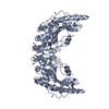
| ||||||||
| 2 | 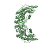
| ||||||||
| 3 | 
| ||||||||
| 4 | 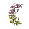
| ||||||||
| Unit cell |
|
-Components
| #1: Protein | Mass: 43170.137 Da / Num. of mol.: 4 / Source method: isolated from a natural source / Source: (natural) Homo sapiens (human) / Tissue: MUSCLE / References: UniProt: P06732, creatine kinase |
|---|
-Experimental details
-Experiment
| Experiment | Method: X-RAY DIFFRACTION / Number of used crystals: 1 |
|---|
- Sample preparation
| Crystal | Density Matthews: 2.69 Å3/Da / Density % sol: 54.23 % | ||||||||||||||||||||||||
|---|---|---|---|---|---|---|---|---|---|---|---|---|---|---|---|---|---|---|---|---|---|---|---|---|---|
| Crystal grow | Temperature: 291 K / Method: vapor diffusion, hanging drop / pH: 8 Details: MPD, Tris-HCL, pH 8.0, VAPOR DIFFUSION, HANGING DROP, temperature 291K | ||||||||||||||||||||||||
| Crystal grow | *PLUS pH: 5.8 / Details: Tang, L., (1998) Acta Crystallogr., D55, 669. | ||||||||||||||||||||||||
| Components of the solutions | *PLUS
|
-Data collection
| Diffraction | Mean temperature: 291 K |
|---|---|
| Diffraction source | Source: SYNCHROTRON / Site: Photon Factory  / Beamline: BL-6A / Wavelength: 1 Å / Beamline: BL-6A / Wavelength: 1 Å |
| Detector | Type: WEISSENBERG / Detector: DIFFRACTOMETER |
| Radiation | Protocol: SINGLE WAVELENGTH / Monochromatic (M) / Laue (L): M / Scattering type: x-ray |
| Radiation wavelength | Wavelength: 1 Å / Relative weight: 1 |
| Reflection | Resolution: 3.5→30 Å / Num. obs: 21262 / % possible obs: 0.883 % / Redundancy: 3 % / Biso Wilson estimate: 24.347 Å2 / Rmerge(I) obs: 0.118 |
| Reflection shell | Resolution: 3.5→3.62 Å / Redundancy: 2 % / Rmerge(I) obs: 0.344 / Mean I/σ(I) obs: 2 / Num. unique all: 2232 / % possible all: 0.935 |
| Reflection | *PLUS Lowest resolution: 30 Å / Num. measured all: 63933 |
| Reflection shell | *PLUS % possible obs: 93.5 % |
- Processing
| Software |
| ||||||||||||||||||||
|---|---|---|---|---|---|---|---|---|---|---|---|---|---|---|---|---|---|---|---|---|---|
| Refinement | Method to determine structure: MOLECULAR REPLACEMENT Starting model: rabbit muscle creatine kinase Resolution: 3.5→8 Å / Rfactor Rfree error: 0.007 / Data cutoff high absF: 4033176.82 / Data cutoff low absF: 0 / Isotropic thermal model: RESTRAINED / Cross valid method: THROUGHOUT / σ(F): 0 / σ(I): 0
| ||||||||||||||||||||
| Solvent computation | Solvent model: FLAT MODEL / Bsol: 107.8 Å2 / ksol: 0.451 e/Å3 | ||||||||||||||||||||
| Displacement parameters | Biso mean: 41.4 Å2
| ||||||||||||||||||||
| Refine analyze |
| ||||||||||||||||||||
| Refinement step | Cycle: LAST / Resolution: 3.5→8 Å
| ||||||||||||||||||||
| Refine LS restraints |
| ||||||||||||||||||||
| Refine LS restraints NCS | NCS model details: CONSTR | ||||||||||||||||||||
| LS refinement shell | Resolution: 3.5→3.7 Å / Rfactor Rfree error: 0.02 / Total num. of bins used: 6
| ||||||||||||||||||||
| Xplor file |
| ||||||||||||||||||||
| Refinement | *PLUS Highest resolution: 3.5 Å / Lowest resolution: 8 Å / Rfactor Rfree: 0.286 | ||||||||||||||||||||
| Solvent computation | *PLUS | ||||||||||||||||||||
| Displacement parameters | *PLUS | ||||||||||||||||||||
| Refine LS restraints | *PLUS
|
