 Movie
Movie Controller

[English] 日本語
 Yorodumi
- PDB-4upa: Crystal structure of Entamoeba histolytica lysyl-tRNA synthetase ... -
+ Open data

- Basic information
| Entry | Database: PDB / ID: 4upa | ||||||
|---|---|---|---|---|---|---|---|
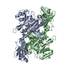
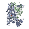
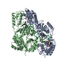
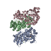
| Title | Crystal structure of Entamoeba histolytica lysyl-tRNA synthetase in complex with AMPPNP | ||||||
 Components Components | LYSINE--TRNA LIGASE | ||||||
 Keywords Keywords | LIGASE / AMINOACYLATION | ||||||
| Function / homology |  Function and homology information Function and homology informationlysine-tRNA ligase / lysine-tRNA ligase activity / lysyl-tRNA aminoacylation / tRNA binding / ATP binding / cytoplasm Similarity search - Function | ||||||
| Biological species |   ENTAMOEBA HISTOLYTICA (eukaryote) ENTAMOEBA HISTOLYTICA (eukaryote) | ||||||
| Method |  X-RAY DIFFRACTION / SYNCHROTRON / MOLECULAR REPLACEMENT / Resolution: 2.901 Å X-RAY DIFFRACTION / SYNCHROTRON / MOLECULAR REPLACEMENT / Resolution: 2.901 Å | ||||||
 Authors Authors | Bonnefond, L. / Nureki, O. | ||||||
 Citation Citation | Journal: FEBS Lett. / Year: 2014 Title: Crystal Structures of Entamoeba Histolytica Lysyl-tRNA Synthetase Reveal Conformational Changes Upon Lysine Binding and a Specific Helix Bundle Domain. Authors: Bonnefond, L. / Castro De Moura, M. / Ribas De Pouplana, L. / Nureki, O. | ||||||
| History |
| ||||||
| Remark 700 | SHEET DETERMINATION METHOD: DSSP THE SHEETS PRESENTED AS "AA" IN EACH CHAIN ON SHEET RECORDS BELOW ... SHEET DETERMINATION METHOD: DSSP THE SHEETS PRESENTED AS "AA" IN EACH CHAIN ON SHEET RECORDS BELOW IS ACTUALLY AN 6-STRANDED BARREL THIS IS REPRESENTED BY A 7-STRANDED SHEET IN WHICH THE FIRST AND LAST STRANDS ARE IDENTICAL. |
- Structure visualization
| Structure viewer | Molecule: MolmilJmol/JSmol |
|---|
- Downloads & links
-Download
| PDBx/mmCIF format | 4upa.cif.gz | 227 KB | Display | PDBx/mmCIF format |
|---|---|---|---|---|
| PDB format | pdb4upa.ent.gz | 179.5 KB | Display | PDB format |
| PDBx/mmJSON format | 4upa.json.gz | Tree view | PDBx/mmJSON format | |
| Others |  Other downloads Other downloads |
-Validation report
| Arichive directory | https://data.pdbj.org/pub/pdb/validation_reports/up/4upaftp://data.pdbj.org/pub/pdb/validation_reports/up/4upa | HTTPS FTP |
|---|
-Related structure data
| Related structure data | 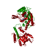 4up7C 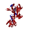 4up8C 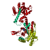 4up9C 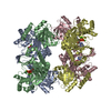 3bjuS C: citing same article ( S: Starting model for refinement |
|---|---|
| Similar structure data |


-Links
 PDBj
PDBj


- Assembly
| Deposited unit | 
| ||||||||
|---|---|---|---|---|---|---|---|---|---|
| 1 | 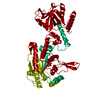
| ||||||||
| Unit cell |
|
-Components
| #1: Protein | Mass: 87757.242 Da / Num. of mol.: 1 / Mutation: YES Source method: isolated from a genetically manipulated source Source: (gene. exp.) ENTAMOEBA HISTOLYTICA (eukaryote) / Plasmid: PET-30 EK/LIC / Production host:  |
|---|---|
| #2: Chemical | ChemComp-ANP /   Mass: 506.196 Da / Num. of mol.: 1 / Source method: obtained synthetically / Formula: C10H17N6O12P3 / Comment: AMP-PNP, energy-carrying molecule analogue*YM Mass: 506.196 Da / Num. of mol.: 1 / Source method: obtained synthetically / Formula: C10H17N6O12P3 / Comment: AMP-PNP, energy-carrying molecule analogue*YM |
-Experimental details
-Experiment
| Experiment | Method: X-RAY DIFFRACTION / Number of used crystals: 1 |
|---|
- Sample preparation
| Crystal | Density Matthews: 3.8 Å3/Da / Density % sol: 67.6 % / Description: NONE |
|---|---|
| Crystal grow | pH: 7.5 Details: 50 MM HEPES PH 8.0, 50 MM NACL, 7.5% PEG 4000, 1.2 MM SPERMINE |
-Data collection
| Diffraction | Mean temperature: 100 K |
|---|---|
| Diffraction source | Source: SYNCHROTRON / Site: Photon Factory  / Beamline: AR-NE3A / Wavelength: 1 / Beamline: AR-NE3A / Wavelength: 1 |
| Detector | Type: ADSC Q270 / Detector: CCD / Date: Dec 17, 2010 |
| Radiation | Protocol: SINGLE WAVELENGTH / Monochromatic (M) / Laue (L): M / Scattering type: x-ray |
| Radiation wavelength | Wavelength: 1 Å / Relative weight: 1 |
| Reflection | Resolution: 2.9→39.2 Å / Num. obs: 29250 / % possible obs: 99.7 % / Observed criterion σ(I): 1 / Redundancy: 6.1 % / Biso Wilson estimate: 84.21 Å2 / Rmerge(I) obs: 0.11 / Net I/σ(I): 15.2 |
| Reflection shell | Resolution: 2.9→3.1 Å / Redundancy: 5.1 % / Mean I/σ(I) obs: 1.1 / % possible all: 99 |
- Processing
| Software |
| |||||||||||||||||||||||||||||||||||||||||||||||||||||||||||||||||||||||||||||
|---|---|---|---|---|---|---|---|---|---|---|---|---|---|---|---|---|---|---|---|---|---|---|---|---|---|---|---|---|---|---|---|---|---|---|---|---|---|---|---|---|---|---|---|---|---|---|---|---|---|---|---|---|---|---|---|---|---|---|---|---|---|---|---|---|---|---|---|---|---|---|---|---|---|---|---|---|---|---|
| Refinement | Method to determine structure: MOLECULAR REPLACEMENT Starting model: PDB ENTRY 3BJU Resolution: 2.901→38.683 Å / SU ML: 0.46 / σ(F): 1.35 / Phase error: 24.82 / Stereochemistry target values: ML
| |||||||||||||||||||||||||||||||||||||||||||||||||||||||||||||||||||||||||||||
| Solvent computation | Shrinkage radii: 0.9 Å / VDW probe radii: 1.11 Å / Solvent model: FLAT BULK SOLVENT MODEL | |||||||||||||||||||||||||||||||||||||||||||||||||||||||||||||||||||||||||||||
| Displacement parameters | Biso mean: 80.7 Å2 | |||||||||||||||||||||||||||||||||||||||||||||||||||||||||||||||||||||||||||||
| Refinement step | Cycle: LAST / Resolution: 2.901→38.683 Å
| |||||||||||||||||||||||||||||||||||||||||||||||||||||||||||||||||||||||||||||
| Refine LS restraints |
| |||||||||||||||||||||||||||||||||||||||||||||||||||||||||||||||||||||||||||||
| LS refinement shell |
| |||||||||||||||||||||||||||||||||||||||||||||||||||||||||||||||||||||||||||||
| Refinement TLS params. | Method: refined / Origin x: 147.0406 Å / Origin y: 126.8564 Å / Origin z: 15.5773 Å
| |||||||||||||||||||||||||||||||||||||||||||||||||||||||||||||||||||||||||||||
| Refinement TLS group | Selection details: ALL |
