[English] 日本語
Yorodumi
- PDB-2ck2: Structure of core-swapped mutant of fibronectin -

+
Open data


ID or keywords:

Loading...

-
Basic information

Entry
Database: PDB / ID: 2ck2
TitleStructure of core-swapped mutant of fibronectin
ComponentsHUMAN FIBRONECTIN
KeywordsSIGNALING PROTEIN / SULFATION / ACUTE PHASE / GLYCOPROTEIN / CELL ADHESION / PYRROLIDONE CARBOXYLIC ACID / HEPARIN-BINDING / PHOSPHORYLATION / ALTERNATIVE SPLICING
Function / homology
Function and homology information


negative regulation of monocyte activation / calcium-independent cell-matrix adhesion / negative regulation of transforming growth factor beta production / Fibronectin matrix formation / Extracellular matrix organization / positive regulation of substrate-dependent cell migration, cell attachment to substrate / neural crest cell migration involved in autonomic nervous system development / peptidase activator activity / fibrinogen complex / peptide cross-linking ...negative regulation of monocyte activation / calcium-independent cell-matrix adhesion / negative regulation of transforming growth factor beta production / Fibronectin matrix formation / Extracellular matrix organization / positive regulation of substrate-dependent cell migration, cell attachment to substrate / neural crest cell migration involved in autonomic nervous system development / peptidase activator activity / fibrinogen complex / peptide cross-linking / integrin activation / ALK mutants bind TKIs / cell-substrate junction assembly / biological process involved in interaction with symbiont / proteoglycan binding / Molecules associated with elastic fibres / extracellular matrix structural constituent / MET activates PTK2 signaling / Syndecan interactions / p130Cas linkage to MAPK signaling for integrins / endodermal cell differentiation / GRB2:SOS provides linkage to MAPK signaling for Integrins / endoplasmic reticulum-Golgi intermediate compartment / Non-integrin membrane-ECM interactions / basement membrane / ECM proteoglycans / Integrin cell surface interactions / positive regulation of axon extension / collagen binding / Degradation of the extracellular matrix / Integrin signaling / substrate adhesion-dependent cell spreading / extracellular matrix / regulation of ERK1 and ERK2 cascade / cell-matrix adhesion / platelet alpha granule lumen / acute-phase response / integrin-mediated signaling pathway / Cell surface interactions at the vascular wall / Post-translational protein phosphorylation / regulation of protein phosphorylation / wound healing / Signaling by high-kinase activity BRAF mutants / MAP2K and MAPK activation / response to wounding / Signaling by RAF1 mutants / Signaling by moderate kinase activity BRAF mutants / Paradoxical activation of RAF signaling by kinase inactive BRAF / Signaling downstream of RAS mutants / Regulation of Insulin-like Growth Factor (IGF) transport and uptake by Insulin-like Growth Factor Binding Proteins (IGFBPs) / GPER1 signaling / positive regulation of fibroblast proliferation / Signaling by BRAF and RAF1 fusions / Signaling by ALK fusions and activated point mutants / integrin binding / Platelet degranulation / nervous system development / heart development / heparin binding / regulation of cell shape / protease binding / Interleukin-4 and Interleukin-13 signaling / angiogenesis / collagen-containing extracellular matrix / blood microparticle / positive regulation of phosphatidylinositol 3-kinase/protein kinase B signal transduction / cell adhesion / apical plasma membrane / endoplasmic reticulum lumen / signaling receptor binding / positive regulation of cell population proliferation / positive regulation of gene expression / extracellular space / extracellular exosome / extracellular region / identical protein binding / plasma membrane
Similarity search - Function
: / Fibronectin type I domain / Fibronectin, type I / Fibronectin type-I domain signature. / Fibronectin type-I domain profile. / Fibronectin type 1 domain / Fibronectin type II domain / Fibronectin type II domain superfamily / Fibronectin type II domain / Fibronectin type-II collagen-binding domain signature. ...: / Fibronectin type I domain / Fibronectin, type I / Fibronectin type-I domain signature. / Fibronectin type-I domain profile. / Fibronectin type 1 domain / Fibronectin type II domain / Fibronectin type II domain superfamily / Fibronectin type II domain / Fibronectin type-II collagen-binding domain signature. / Fibronectin type-II collagen-binding domain profile. / Fibronectin type 2 domain / Kringle-like fold / EGF-like domain signature 1. / Fibronectin type III domain / Fibronectin type 3 domain / Fibronectin type-III domain profile. / Fibronectin type III / Fibronectin type III superfamily / Immunoglobulins / Immunoglobulin-like fold / Immunoglobulin-like / Sandwich / Mainly Beta
Similarity search - Domain/homology
ACETYL GROUP / Fibronectin
Similarity search - Component
Biological speciesHOMO SAPIENS (human)
MethodX-RAY DIFFRACTION / MOLECULAR REPLACEMENT / Resolution: 2 Å
AuthorsNg, S.P. / Billings, K.S. / Ohashi, T. / Allen, M.D. / Best, R.B. / Randles, L.G. / Erickson, H.P. / Clarke, J.
CitationJournal: Proc.Natl.Acad.Sci.USA / Year: 2007
Title: Designing an Extracellular Matrix Protein with Enhanced Mechanical Stability
Authors: Ng, S.P. / Billings, K.S. / Ohashi, T. / Allen, M.D. / Best, R.B. / Randles, L.G. / Erickson, H.P. / Clarke, J.
History
DepositionApr 10, 2006Deposition site: PDBE / Processing site: PDBE
Revision 1.0Apr 10, 2007Provider: repository / Type: Initial release
Revision 1.1Apr 15, 2015Group: Database references / Derived calculations ...Database references / Derived calculations / Other / Version format compliance
Revision 1.2Jan 24, 2018Group: Source and taxonomy / Category: entity_src_gen
Item: _entity_src_gen.pdbx_host_org_ncbi_taxonomy_id / _entity_src_gen.pdbx_host_org_scientific_name ..._entity_src_gen.pdbx_host_org_ncbi_taxonomy_id / _entity_src_gen.pdbx_host_org_scientific_name / _entity_src_gen.pdbx_host_org_strain / _entity_src_gen.pdbx_host_org_variant
Revision 1.3Dec 13, 2023Group: Data collection / Database references ...Data collection / Database references / Derived calculations / Other / Refinement description
Category: chem_comp_atom / chem_comp_bond ...chem_comp_atom / chem_comp_bond / database_2 / pdbx_database_status / pdbx_initial_refinement_model / struct_site
Item: _database_2.pdbx_DOI / _database_2.pdbx_database_accession ..._database_2.pdbx_DOI / _database_2.pdbx_database_accession / _pdbx_database_status.status_code_sf / _struct_site.pdbx_auth_asym_id / _struct_site.pdbx_auth_comp_id / _struct_site.pdbx_auth_seq_id
Remark 700 
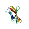
SHEET THE SHEET STRUCTURE OF THIS MOLECULE IS BIFURCATED. IN ORDER TO REPRESENT THIS FEATURE IN ... SHEET THE SHEET STRUCTURE OF THIS MOLECULE IS BIFURCATED. IN ORDER TO REPRESENT THIS FEATURE IN THE SHEET RECORDS BELOW, TWO SHEETS ARE DEFINED.

-
Structure visualization

Structure viewerMolecule:
MolmilJmol/JSmol

Downloads & links

-
Assembly

Deposited unit
A: HUMAN FIBRONECTIN
B: HUMAN FIBRONECTIN
hetero molecules


Theoretical massNumber of molelcules
Total (without water)20,4314
Polymers20,3432
Non-polymers882
Water1,964109
1


  • Idetical with deposited unit
  • defined by author&software
TypeNameSymmetry operationNumber
identity operation1_555x,y,z1
MethodPQS
Unit cell
Length a, b, c (Å)36.703, 37.227, 137.834
Angle α, β, γ (deg.)90.00, 90.00, 90.00
Int Tables number19
Space group name H-MP212121

-
Components

#1: Protein HUMAN FIBRONECTIN / FN / COLD-INSOLUBLE GLOBULIN / CIG


Mass: 10171.315 Da / Num. of mol.: 2 / Fragment: RESIDUES 1447-1542 / Mutation: YES
Source method: isolated from a genetically manipulated source
Source: (gene. exp.) HOMO SAPIENS (human) / Production host: ESCHERICHIA COLI BL21(DE3) (bacteria) / Variant (production host): C41 / References: UniProt: P02751
#2: Chemical ChemComp-ACE / ACETYL GROUP


Mass: 44.053 Da / Num. of mol.: 2 / Source method: obtained synthetically / Formula: C2H4O
#3: Water ChemComp-HOH / water


Mass: 18.015 Da / Num. of mol.: 109 / Source method: isolated from a natural source / Formula: H2O
Compound details
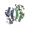
ENGINEERED RESIDUE IN CHAIN A, LEU 1454 TO ILE ENGINEERED RESIDUE IN CHAIN A, ALA 1459 TO VAL ...ENGINEERED RESIDUE IN CHAIN A, LEU 1454 TO ILE ENGINEERED RESIDUE IN CHAIN A, ALA 1459 TO VAL ENGINEERED RESIDUE IN CHAIN A, LEU 1464 TO ALA ENGINEERED RESIDUE IN CHAIN A, VAL 1475 TO ILE ENGINEERED RESIDUE IN CHAIN A, TYR 1478 TO ILE ENGINEERED RESIDUE IN CHAIN A, ILE 1480 TO LEU ENGINEERED RESIDUE IN CHAIN A, PHE 1494 TO ILE ENGINEERED RESIDUE IN CHAIN A, VAL 1496 TO LEU ENGINEERED RESIDUE IN CHAIN A, ALA 1503 TO TYR ENGINEERED RESIDUE IN CHAIN A, VAL 1512 TO THR ENGINEERED RESIDUE IN CHAIN A, VAL 1518 TO LEU ENGINEERED RESIDUE IN CHAIN A, ALA 1520 TO SER ENGINEERED RESIDUE IN CHAIN A, ILE 1534 TO ALA ENGINEERED RESIDUE IN CHAIN A, TYR 1538 TO PHE ENGINEERED RESIDUE IN CHAIN B, LEU 1454 TO ILE ENGINEERED RESIDUE IN CHAIN B, ALA 1459 TO VAL ENGINEERED RESIDUE IN CHAIN B, LEU 1464 TO ALA ENGINEERED RESIDUE IN CHAIN B, VAL 1475 TO ILE ENGINEERED RESIDUE IN CHAIN B, TYR 1478 TO ILE ENGINEERED RESIDUE IN CHAIN B, ILE 1480 TO LEU ENGINEERED RESIDUE IN CHAIN B, PHE 1494 TO ILE ENGINEERED RESIDUE IN CHAIN B, VAL 1496 TO LEU ENGINEERED RESIDUE IN CHAIN B, ALA 1503 TO TYR ENGINEERED RESIDUE IN CHAIN B, VAL 1512 TO THR ENGINEERED RESIDUE IN CHAIN B, VAL 1518 TO LEU ENGINEERED RESIDUE IN CHAIN B, ALA 1520 TO SER ENGINEERED RESIDUE IN CHAIN B, ILE 1534 TO ALA ENGINEERED RESIDUE IN CHAIN B, TYR 1538 TO PHE
Sequence detailsDELIBERATELY ENGINEERED MUTATION

-
Experimental details

-
Experiment

ExperimentMethod: X-RAY DIFFRACTION / Number of used crystals: 1

-
Sample preparation

CrystalDensity Matthews: 2.32 Å3/Da / Density % sol: 47 %
Crystal growpH: 5.1
Details: 50% PEG-400, 0.1 M ACTEATE, 0.2 M LITHIUM SULPHATE, PH 5.1

-
Data collection

DiffractionMean temperature: 100 K
Diffraction sourceSource: ROTATING ANODE / Wavelength: 1.5418
DetectorDate: Feb 2, 2006
RadiationProtocol: SINGLE WAVELENGTH / Monochromatic (M) / Laue (L): M / Scattering type: x-ray
Radiation wavelengthWavelength: 1.5418 Å / Relative weight: 1
ReflectionResolution: 2→19.55 Å / Num. obs: 12995 / % possible obs: 97.3 % / Observed criterion σ(I): 1 / Redundancy: 8.9 % / Rmerge(I) obs: 0.07 / Net I/σ(I): 23
Reflection shellResolution: 2→2.11 Å / Redundancy: 8.5 % / Rmerge(I) obs: 0.23 / Mean I/σ(I) obs: 8.9 / % possible all: 95.9

-
Processing

Software
NameVersionClassification
CNS1.1refinement
MOSFLMdata reduction
SCALAdata scaling
PHASERphasing
RefinementMethod to determine structure: MOLECULAR REPLACEMENT
Starting model: PDB ENTRY 1TEN
Resolution: 2→19.55 Å / Data cutoff high absF: 10000 / Cross valid method: THROUGHOUT / σ(F): 0 / Stereochemistry target values: TWIN_LSQ
Details: COORDINATES FROM TWINNED DATA RESTRAINED INDIVIDUAL B-REFINEMENT TWINNING OPERATOR K,H,-L TWINNING FRACTION 0.185
RfactorNum. reflection% reflectionSelection details
Rfree0.2683 634 4.7 %RANDOM
Rwork0.2008 ---
obs0.2008 12656 94.3 %-
Solvent computationSolvent model: DENSITY MODIFICATION / Bsol: 58.6329 Å2 / ksol: 0.386987 e/Å3
Displacement parameters
Baniso -1Baniso -2Baniso -3
1--2.17 Å20 Å20 Å2
2---0.142 Å20 Å2
3---2.312 Å2
Refinement stepCycle: LAST / Resolution: 2→19.55 Å
ProteinNucleic acidLigandSolventTotal
Num. atoms1420 0 6 109 1535
Refine LS restraints
Refine-IDTypeDev ideal
X-RAY DIFFRACTIONc_bond_d0.008
X-RAY DIFFRACTIONc_bond_d_na
X-RAY DIFFRACTIONc_bond_d_prot
X-RAY DIFFRACTIONc_angle_d
X-RAY DIFFRACTIONc_angle_d_na
X-RAY DIFFRACTIONc_angle_d_prot
X-RAY DIFFRACTIONc_angle_deg1.5
X-RAY DIFFRACTIONc_angle_deg_na
X-RAY DIFFRACTIONc_angle_deg_prot
X-RAY DIFFRACTIONc_dihedral_angle_d
X-RAY DIFFRACTIONc_dihedral_angle_d_na
X-RAY DIFFRACTIONc_dihedral_angle_d_prot
X-RAY DIFFRACTIONc_improper_angle_d
X-RAY DIFFRACTIONc_improper_angle_d_na
X-RAY DIFFRACTIONc_improper_angle_d_prot
X-RAY DIFFRACTIONc_mcbond_it
X-RAY DIFFRACTIONc_mcangle_it
X-RAY DIFFRACTIONc_scbond_it
X-RAY DIFFRACTIONc_scangle_it
Xplor file
Refine-IDSerial noParam file
X-RAY DIFFRACTION1PROTEIN_REP.PARAM
X-RAY DIFFRACTION2WATER_REP.PARAM
X-RAY DIFFRACTION3ACE.PAR

+
About Yorodumi

-
News

-
Feb 9, 2022. New format data for meta-information of EMDB entries

New format data for meta-information of EMDB entries

  • Version 3 of the EMDB header file is now the official format.
  • The previous official version 1.9 will be removed from the archive.

Related info.:EMDB header

External links:wwPDB to switch to version 3 of the EMDB data model

-
Aug 12, 2020. Covid-19 info

Covid-19 info

URL: https://pdbjlc1.pdbj.org/emnavi/covid19.php

New page: Covid-19 featured information page in EM Navigator.

Related info.:Covid-19 info / Mar 5, 2020. Novel coronavirus structure data

+
Mar 5, 2020. Novel coronavirus structure data

Novel coronavirus structure data

Related info.:Yorodumi Speices / Aug 12, 2020. Covid-19 info

External links:COVID-19 featured content - PDBj / Molecule of the Month (242):Coronavirus Proteases

+
Jan 31, 2019. EMDB accession codes are about to change! (news from PDBe EMDB page)

EMDB accession codes are about to change! (news from PDBe EMDB page)

  • The allocation of 4 digits for EMDB accession codes will soon come to an end. Whilst these codes will remain in use, new EMDB accession codes will include an additional digit and will expand incrementally as the available range of codes is exhausted. The current 4-digit format prefixed with “EMD-” (i.e. EMD-XXXX) will advance to a 5-digit format (i.e. EMD-XXXXX), and so on. It is currently estimated that the 4-digit codes will be depleted around Spring 2019, at which point the 5-digit format will come into force.
  • The EM Navigator/Yorodumi systems omit the EMD- prefix.

Related info.:Q: What is EMD? / ID/Accession-code notation in Yorodumi/EM Navigator

External links:EMDB Accession Codes are Changing Soon! / Contact to PDBj

+
Jul 12, 2017. Major update of PDB

Major update of PDB

  • wwPDB released updated PDB data conforming to the new PDBx/mmCIF dictionary.
  • This is a major update changing the version number from 4 to 5, and with Remediation, in which all the entries are updated.
  • In this update, many items about electron microscopy experimental information are reorganized (e.g. em_software).
  • Now, EM Navigator and Yorodumi are based on the updated data.

External links:wwPDB Remediation / Enriched Model Files Conforming to OneDep Data Standards Now Available in the PDB FTP Archive

-
Yorodumi

Thousand views of thousand structures

  • Yorodumi is a browser for structure data from EMDB, PDB, SASBDB, etc.
  • This page is also the successor to EM Navigator detail page, and also detail information page/front-end page for Omokage search.
  • The word "yorodu" (or yorozu) is an old Japanese word meaning "ten thousand". "mi" (miru) is to see.

Related info.:EMDB / PDB / SASBDB / Comparison of 3 databanks / Yorodumi Search / Aug 31, 2016. New EM Navigator & Yorodumi / Yorodumi Papers / Jmol/JSmol / Function and homology information / Changes in new EM Navigator and Yorodumi

Read more