 Movie
Movie Controller

[English] 日本語
 Yorodumi
- PDB-2cjw: Crystal structure of the small GTPase Gem (GemDNDCaM) in complex ... -
+ Open data

- Basic information
| Entry | Database: PDB / ID: 2cjw | ||||||
|---|---|---|---|---|---|---|---|
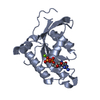
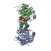
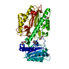
| Title | Crystal structure of the small GTPase Gem (GemDNDCaM) in complex to Mg.GDP | ||||||
 Components Components | (GTP-BINDING PROTEIN GEM) x 2 | ||||||
 Keywords Keywords | G-PROTEIN / NUCLEOTIDE-BINDING / GTP-BINDING / SMALL GTPASE / CONFORMATIONAL CHANGE / CYSTEINE-MODIFIED / G-PROTEIN HYDROLASE | ||||||
| Function / homology |  Function and homology information Function and homology informationmetaphase chromosome alignment / spindle midzone / chromosome organization / NPAS4 regulates expression of target genes / calcium channel regulator activity / cytoplasmic side of plasma membrane / mitotic spindle / GDP binding / mitotic cell cycle / midbody ...metaphase chromosome alignment / spindle midzone / chromosome organization / NPAS4 regulates expression of target genes / calcium channel regulator activity / cytoplasmic side of plasma membrane / mitotic spindle / GDP binding / mitotic cell cycle / midbody / calmodulin binding / cell surface receptor signaling pathway / immune response / GTPase activity / GTP binding / magnesium ion binding / signal transduction / nucleus / plasma membrane Similarity search - Function | ||||||
| Biological species |  HOMO SAPIENS (human) HOMO SAPIENS (human) | ||||||
| Method |  X-RAY DIFFRACTION / SYNCHROTRON / MOLECULAR REPLACEMENT / Resolution: 2.1 Å X-RAY DIFFRACTION / SYNCHROTRON / MOLECULAR REPLACEMENT / Resolution: 2.1 Å | ||||||
 Authors Authors | Splingard, A. / Menetrey, J. / Perderiset, M. / Cicolari, J. / Hamoudi, F. / Cabanie, L. / El Marjou, A. / Wells, A. / Houdusse, A. / de Gunzburg, J. | ||||||
 Citation Citation | Journal: J.Biol.Chem. / Year: 2007 Title: Biochemical and Structural Characterization of the Gem Gtpase. Authors: Splingard, A. / Menetrey, J. / Perderiset, M. / Cicolari, J. / Regazzoni, K. / Hamoudi, F. / Cabanie, L. / El Marjou, A. / Wells, A. / Houdusse, A. / De Gunzburg, J. | ||||||
| History |
|
- Structure visualization
| Structure viewer | Molecule: MolmilJmol/JSmol |
|---|
- Downloads & links
-Download
| PDBx/mmCIF format | 2cjw.cif.gz | 92.1 KB | Display | PDBx/mmCIF format |
|---|---|---|---|---|
| PDB format | pdb2cjw.ent.gz | 68.6 KB | Display | PDB format |
| PDBx/mmJSON format | 2cjw.json.gz | Tree view | PDBx/mmJSON format | |
| Others |  Other downloads Other downloads |
-Validation report
| Arichive directory | https://data.pdbj.org/pub/pdb/validation_reports/cj/2cjwftp://data.pdbj.org/pub/pdb/validation_reports/cj/2cjw | HTTPS FTP |
|---|
-Related structure data
| Related structure data | 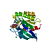 1kaoS S: Starting model for refinement |
|---|---|
| Similar structure data |


-Links
 PDBj
PDBj

- Assembly
| Deposited unit | 
| ||||||||
|---|---|---|---|---|---|---|---|---|---|
| 1 |
| ||||||||
| Unit cell |
|
-Components
| #1: Protein | Mass: 22431.904 Da / Num. of mol.: 1 / Fragment: G DOMAIN, RESIDUES 74-261 Source method: isolated from a genetically manipulated source Source: (gene. exp.) HOMO SAPIENS (human) / Production host:  | ||||||||
|---|---|---|---|---|---|---|---|---|---|
| #2: Protein | Mass: 22431.904 Da / Num. of mol.: 1 / Fragment: G DOMAIN, RESIDUES 74-261 Source method: isolated from a genetically manipulated source Source: (gene. exp.) HOMO SAPIENS (human) / Production host: | ||||||||
| #3: Chemical |   Type: RNA linking / Mass: 443.201 Da / Num. of mol.: 2 / Source method: obtained synthetically / Formula: C10H15N5O11P2 / Comment: GDP, energy-carrying molecule*YM Type: RNA linking / Mass: 443.201 Da / Num. of mol.: 2 / Source method: obtained synthetically / Formula: C10H15N5O11P2 / Comment: GDP, energy-carrying molecule*YM#4: Chemical |   Mass: 24.305 Da / Num. of mol.: 2 / Source method: obtained synthetically / Formula: Mg Mass: 24.305 Da / Num. of mol.: 2 / Source method: obtained synthetically / Formula: Mg#5: Water | ChemComp-HOH / |  Mass: 18.015 Da / Num. of mol.: 127 / Source method: isolated from a natural source / Formula: H2O Mass: 18.015 Da / Num. of mol.: 127 / Source method: isolated from a natural source / Formula: H2OHas protein modification | Y | Sequence details | RESIDUES 70-72 (EFG) IN MOLECULE B BELONG TO THE REMAINING PART OF THE TAG. | |
-Experimental details
-Experiment
| Experiment | Method: X-RAY DIFFRACTION |
|---|
- Sample preparation
| Crystal | Density Matthews: 3.65 Å3/Da / Density % sol: 66.3 % |
|---|---|
| Crystal grow | Details: 0.1M NACACODYLATE PH6.5, 0.9M NAACETATE, 5MM MGCL2 AND 2MM DTT |
-Data collection
| Diffraction | Mean temperature: 100 K |
|---|---|
| Diffraction source | Source: SYNCHROTRON / Site: ESRF  / Beamline: ID14-1 / Wavelength: 0.96115 / Beamline: ID14-1 / Wavelength: 0.96115 |
| Detector | Type: MARRESEARCH / Detector: CCD / Date: Sep 8, 2004 |
| Radiation | Protocol: SINGLE WAVELENGTH / Monochromatic (M) / Laue (L): M / Scattering type: x-ray |
| Radiation wavelength | Wavelength: 0.96115 Å / Relative weight: 1 |
| Reflection | Resolution: 2.1→60 Å / Num. obs: 36826 / % possible obs: 100 % / Observed criterion σ(I): 2 / Redundancy: 7.7 % / Rmerge(I) obs: 0.086 / Net I/σ(I): 19 |
| Reflection shell | Resolution: 2.1→2.21 Å / Redundancy: 7.7 % / Rmerge(I) obs: 0.342 / Mean I/σ(I) obs: 5.3 / % possible all: 99.9 |
- Processing
| Software |
| ||||||||||||||||||||||||||||||||||||||||||||||||||||||||||||||||||||||||||||||||||||||||||||||||||||||||||||||||||||||||||||||||||||||||||||||||||||||||||||||||||||||||||||||||||||||
|---|---|---|---|---|---|---|---|---|---|---|---|---|---|---|---|---|---|---|---|---|---|---|---|---|---|---|---|---|---|---|---|---|---|---|---|---|---|---|---|---|---|---|---|---|---|---|---|---|---|---|---|---|---|---|---|---|---|---|---|---|---|---|---|---|---|---|---|---|---|---|---|---|---|---|---|---|---|---|---|---|---|---|---|---|---|---|---|---|---|---|---|---|---|---|---|---|---|---|---|---|---|---|---|---|---|---|---|---|---|---|---|---|---|---|---|---|---|---|---|---|---|---|---|---|---|---|---|---|---|---|---|---|---|---|---|---|---|---|---|---|---|---|---|---|---|---|---|---|---|---|---|---|---|---|---|---|---|---|---|---|---|---|---|---|---|---|---|---|---|---|---|---|---|---|---|---|---|---|---|---|---|---|---|
| Refinement | Method to determine structure: MOLECULAR REPLACEMENT Starting model: PDB ENTRY 1KAO Resolution: 2.1→60 Å / Cor.coef. Fo:Fc: 0.94 / Cor.coef. Fo:Fc free: 0.918 / SU B: 3.452 / SU ML: 0.096 / Cross valid method: THROUGHOUT / ESU R: 0.168 / ESU R Free: 0.156 / Stereochemistry target values: MAXIMUM LIKELIHOOD Details: HYDROGENS HAVE BEEN ADDED IN THE RIDING POSITIONS. RESIDUES 102-105 AND 136-139 IN MOLECULE A AND RESIDUES 99-110 AND 136-142 IN MOLECULE B WERE DISORDERED AND NOT MODELED. SOME SIDE-CHAINS ...Details: HYDROGENS HAVE BEEN ADDED IN THE RIDING POSITIONS. RESIDUES 102-105 AND 136-139 IN MOLECULE A AND RESIDUES 99-110 AND 136-142 IN MOLECULE B WERE DISORDERED AND NOT MODELED. SOME SIDE-CHAINS WERE MODELED WITH 0.0 OR PARTIAL OCCUPANCY RATE.
| ||||||||||||||||||||||||||||||||||||||||||||||||||||||||||||||||||||||||||||||||||||||||||||||||||||||||||||||||||||||||||||||||||||||||||||||||||||||||||||||||||||||||||||||||||||||
| Solvent computation | Ion probe radii: 0.8 Å / Shrinkage radii: 0.8 Å / VDW probe radii: 1.4 Å / Solvent model: MASK | ||||||||||||||||||||||||||||||||||||||||||||||||||||||||||||||||||||||||||||||||||||||||||||||||||||||||||||||||||||||||||||||||||||||||||||||||||||||||||||||||||||||||||||||||||||||
| Displacement parameters | Biso mean: 35.22 Å2
| ||||||||||||||||||||||||||||||||||||||||||||||||||||||||||||||||||||||||||||||||||||||||||||||||||||||||||||||||||||||||||||||||||||||||||||||||||||||||||||||||||||||||||||||||||||||
| Refinement step | Cycle: LAST / Resolution: 2.1→60 Å
| ||||||||||||||||||||||||||||||||||||||||||||||||||||||||||||||||||||||||||||||||||||||||||||||||||||||||||||||||||||||||||||||||||||||||||||||||||||||||||||||||||||||||||||||||||||||
| Refine LS restraints |
|