| Entry | Database: PDB / ID: 2ci8
|
|---|
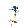
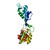
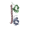
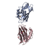
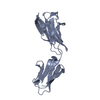
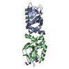
| Title | sh2 domain of human nck1 adaptor protein - uncomplexed |
|---|
 Components Components | CYTOPLASMIC PROTEIN NCK1 |
|---|
 Keywords Keywords | TRANSLATION / BINDING SPECIFICITY / HOST-PATHOGEN / INTERACTIONS |
|---|
| Function / homology |  Function and homology information Function and homology information
positive regulation of cap-dependent translational initiation / regulation of translation initiation in response to endoplasmic reticulum stress / eukaryotic initiation factor eIF2 binding / positive regulation of translation in response to endoplasmic reticulum stress / protein phosphatase type 1 complex / positive regulation of endoplasmic reticulum stress-induced intrinsic apoptotic signaling pathway / positive regulation of cap-independent translational initiation / substrate-dependent cell migration, cell extension / cytoskeletal adaptor activity / signal complex assembly ...positive regulation of cap-dependent translational initiation / regulation of translation initiation in response to endoplasmic reticulum stress / eukaryotic initiation factor eIF2 binding / positive regulation of translation in response to endoplasmic reticulum stress / protein phosphatase type 1 complex / positive regulation of endoplasmic reticulum stress-induced intrinsic apoptotic signaling pathway / positive regulation of cap-independent translational initiation / substrate-dependent cell migration, cell extension / cytoskeletal adaptor activity / signal complex assembly / Activation of RAC1 / DCC mediated attractive signaling / Nephrin family interactions / vesicle membrane / lamellipodium assembly / RHOV GTPase cycle / positive regulation of actin filament polymerization / negative regulation of PERK-mediated unfolded protein response / protein kinase inhibitor activity / RHOU GTPase cycle / Generation of second messenger molecules / ephrin receptor signaling pathway / negative regulation of T cell receptor signaling pathway / RHO GTPases Activate WASPs and WAVEs / ephrin receptor binding / negative regulation of insulin receptor signaling pathway / positive regulation of T cell proliferation / signaling adaptor activity / Downstream signal transduction / regulation of cell migration / response to endoplasmic reticulum stress / Antigen activates B Cell Receptor (BCR) leading to generation of second messengers / antiviral innate immune response / actin filament organization / T cell activation / FCGR3A-mediated phagocytosis / protein sequestering activity / molecular condensate scaffold activity / positive regulation of neuron projection development / receptor tyrosine kinase binding / Regulation of actin dynamics for phagocytic cup formation / PKR-mediated signaling / VEGFA-VEGFR2 Pathway / cell-cell junction / cell migration / signaling receptor complex adaptor activity / Potential therapeutics for SARS / protein-macromolecule adaptor activity / cadherin binding / ribosome / protein domain specific binding / signaling receptor binding / negative regulation of transcription by RNA polymerase II / endoplasmic reticulum / positive regulation of transcription by RNA polymerase II / nucleus / plasma membrane / cytosol / cytoplasmSimilarity search - Function Nck1, SH3 domain 1 / Nck1, SH3 domain 2 / Nck1, SH3 domain 3 / Nck1, SH2 domain / Cytoplasmic protein NCK / : / SH2 domain / SHC Adaptor Protein / SH3 domain / SH2 domain ...Nck1, SH3 domain 1 / Nck1, SH3 domain 2 / Nck1, SH3 domain 3 / Nck1, SH2 domain / Cytoplasmic protein NCK / : / SH2 domain / SHC Adaptor Protein / SH3 domain / SH2 domain / Src homology 2 (SH2) domain profile. / Src homology 2 domains / SH2 domain / Src homology 3 domains / SH2 domain superfamily / SH3-like domain superfamily / Src homology 3 (SH3) domain profile. / SH3 domain / 2-Layer Sandwich / Alpha BetaSimilarity search - Domain/homology |
|---|
| Biological species |  HOMO SAPIENS (human) HOMO SAPIENS (human) |
|---|
| Method |  X-RAY DIFFRACTION / SYNCHROTRON / MOLECULAR REPLACEMENT / Resolution: 1.8 Å X-RAY DIFFRACTION / SYNCHROTRON / MOLECULAR REPLACEMENT / Resolution: 1.8 Å |
|---|
 Authors Authors | Frese, S. / Schubert, W.-D. / Findeis, A.C. / Marquardt, T. / Roske, Y.S. / Stradal, T.E.B. / Heinz, D.W. |
|---|
 Citation Citation | Journal: J.Biol.Chem. / Year: 2006
Title: The Phosphotyrosine Peptide Binding Specificity of Nck1 and Nck2 Src Homology 2 Domains.
Authors: Frese, S. / Schubert, W.-D. / Findeis, A.C. / Marquardt, T. / Roske, Y.S. / Stradal, T.E.B. / Heinz, D.W. |
|---|
| History | | Deposition | Mar 17, 2006 | Deposition site: PDBE / Processing site: PDBE |
|---|
| Revision 1.0 | Apr 24, 2006 | Provider: repository / Type: Initial release |
|---|
| Revision 1.1 | Jul 13, 2011 | Group: Advisory / Version format compliance |
|---|
| Revision 1.2 | Apr 3, 2019 | Group: Advisory / Data collection ...Advisory / Data collection / Derived calculations / Experimental preparation / Other
Category: database_PDB_caveat / database_PDB_rev ...database_PDB_caveat / database_PDB_rev / database_PDB_rev_record / exptl_crystal_grow / pdbx_data_processing_status / pdbx_database_proc / pdbx_database_status / struct_conn / struct_conn_type
Item: _exptl_crystal_grow.temp / _pdbx_database_status.recvd_author_approval |
|---|
| Revision 1.3 | May 15, 2019 | Group: Data collection / Experimental preparation / Category: exptl_crystal_grow / Item: _exptl_crystal_grow.method |
|---|
| Revision 1.4 | Dec 13, 2023 | Group: Data collection / Database references ...Data collection / Database references / Derived calculations / Other / Refinement description
Category: chem_comp_atom / chem_comp_bond ...chem_comp_atom / chem_comp_bond / database_2 / pdbx_database_status / pdbx_initial_refinement_model / struct_site
Item: _database_2.pdbx_DOI / _database_2.pdbx_database_accession ..._database_2.pdbx_DOI / _database_2.pdbx_database_accession / _pdbx_database_status.status_code_sf / _struct_site.pdbx_auth_asym_id / _struct_site.pdbx_auth_comp_id / _struct_site.pdbx_auth_seq_id |
|---|
|
|---|
 Movie
Movie Controller


 Open data
 Basic information
 Structure visualization
 Downloads & links
 Other downloads


 PDBj
PDBj


 Assembly


 Mass: 96.063 Da / Num. of mol.: 4 / Source method: obtained synthetically / Formula: SO4
 Mass: 238.278 Da / Num. of mol.: 1 / Source method: obtained synthetically / Formula: C10H22O6 / Comment: precipitant*YM
 Mass: 18.015 Da / Num. of mol.: 67 / Source method: isolated from a natural source / Formula: H2O
 Sample preparation
 / Beamline: 14.1 / Wavelength: 0.91977
  Processing