| Entry | Database: PDB / ID: 2cfb
|
|---|
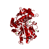
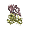
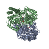
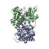
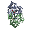
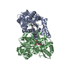
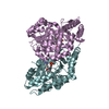
| Title | Glutamate-1-semialdehyde 2,1-Aminomutase from Thermosynechococcus elongatus |
|---|
 Components Components | GLUTAMATE-1-SEMIALDEHYDE 2,1-AMINOMUTASE |
|---|
 Keywords Keywords | ISOMERASE / TETRAPYRROLE BIOSYNTHESIS / PYRIDOXAL PHOSPHATE DEPENDENT / AMINOTRANSFERASE / PORPHYRIN BIOSYNTHESIS / CHLOROPHYLL BIOSYNTHESIS |
|---|
| Function / homology |  Function and homology information Function and homology information
glutamate-1-semialdehyde 2,1-aminomutase / glutamate-1-semialdehyde 2,1-aminomutase activity / chlorophyll biosynthetic process / : / transaminase activity / pyridoxal phosphate binding / cytoplasmSimilarity search - Function Tetrapyrrole biosynthesis, glutamate-1-semialdehyde aminotransferase / : / Aminotransferases class-III pyridoxal-phosphate attachment site. / Aminotransferase class-III / Aminotransferase class-III / Aspartate Aminotransferase, domain 1 / Aspartate Aminotransferase, domain 1 / Aspartate Aminotransferase; domain 2 / Type I PLP-dependent aspartate aminotransferase-like (Major domain) / Pyridoxal phosphate-dependent transferase, small domain ...Tetrapyrrole biosynthesis, glutamate-1-semialdehyde aminotransferase / : / Aminotransferases class-III pyridoxal-phosphate attachment site. / Aminotransferase class-III / Aminotransferase class-III / Aspartate Aminotransferase, domain 1 / Aspartate Aminotransferase, domain 1 / Aspartate Aminotransferase; domain 2 / Type I PLP-dependent aspartate aminotransferase-like (Major domain) / Pyridoxal phosphate-dependent transferase, small domain / Pyridoxal phosphate-dependent transferase, major domain / Pyridoxal phosphate-dependent transferase / Alpha-Beta Complex / 3-Layer(aba) Sandwich / Alpha BetaSimilarity search - Domain/homology |
|---|
| Biological species |  SYNECHOCOCCUS ELONGATUS (bacteria) SYNECHOCOCCUS ELONGATUS (bacteria) |
|---|
| Method |  X-RAY DIFFRACTION / MOLECULAR REPLACEMENT / Resolution: 2.85 Å X-RAY DIFFRACTION / MOLECULAR REPLACEMENT / Resolution: 2.85 Å |
|---|
 Authors Authors | Schulze, J.O. / Schubert, W.-D. / Moser, J. / Jahn, D. / Heinz, D.W. |
|---|
 Citation Citation | Journal: J.Mol.Biol. / Year: 2006
Title: Evolutionary Relationship between Initial Enzymes of Tetrapyrrole Biosynthesis
Authors: Schulze, J.O. / Schubert, W.-D. / Moser, J. / Jahn, D. / Heinz, D.W. |
|---|
| History | | Deposition | Feb 17, 2006 | Deposition site: PDBE / Processing site: PDBE |
|---|
| Revision 1.0 | Mar 29, 2006 | Provider: repository / Type: Initial release |
|---|
| Revision 1.1 | Jul 13, 2011 | Group: Advisory / Version format compliance |
|---|
| Revision 1.2 | Dec 13, 2023 | Group: Data collection / Database references ...Data collection / Database references / Derived calculations / Other / Refinement description
Category: chem_comp_atom / chem_comp_bond ...chem_comp_atom / chem_comp_bond / database_2 / pdbx_database_status / pdbx_initial_refinement_model / struct_conn
Item: _database_2.pdbx_DOI / _database_2.pdbx_database_accession ..._database_2.pdbx_DOI / _database_2.pdbx_database_accession / _pdbx_database_status.status_code_sf / _struct_conn.pdbx_leaving_atom_flag |
|---|
| Revision 1.3 | Nov 13, 2024 | Group: Structure summary / Category: pdbx_entry_details / pdbx_modification_feature / Item: _pdbx_entry_details.has_protein_modification |
|---|
|
|---|
| Remark 650 | HELIX DETERMINATION METHOD: AUTHOR PROVIDED. |
|---|
| Remark 700 | SHEET DETERMINATION METHOD: PROVIDED BY DEPOSITIOR |
|---|
 Movie
Movie Controller

 Yorodumi
 Open data
 Basic information
 Structure visualization
 Downloads & links
 Other downloads


 PDBj
PDBj Assembly

 Mass: 233.158 Da / Num. of mol.: 1 / Source method: obtained synthetically / Formula: C8H12NO5P
 Mass: 18.015 Da / Num. of mol.: 13 / Source method: isolated from a natural source / Formula: H2O
 Sample preparation
 Processing