 Movie
Movie Controller

[English] 日本語
 Yorodumi
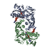
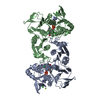
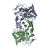
- PDB-2cdu: The Crystal Structure of Water-forming NAD(P)H Oxidase from Lacto... -
+ Open data

- Basic information
| Entry | Database: PDB / ID: 2cdu | |||||||||
|---|---|---|---|---|---|---|---|---|---|---|
| Title | The Crystal Structure of Water-forming NAD(P)H Oxidase from Lactobacillus sanfranciscensis | |||||||||
 Components Components | NADPH OXIDASE | |||||||||
 Keywords Keywords | OXIDOREDUCTASE / NAD(P)H OXIDASE / FLAVOENZYME | |||||||||
| Function / homology |  Function and homology information Function and homology information | |||||||||
| Biological species |  LACTOBACILLUS SANFRANCISCENSIS (bacteria) LACTOBACILLUS SANFRANCISCENSIS (bacteria) | |||||||||
| Method |  X-RAY DIFFRACTION / SYNCHROTRON / MOLECULAR REPLACEMENT / Resolution: 1.8 Å X-RAY DIFFRACTION / SYNCHROTRON / MOLECULAR REPLACEMENT / Resolution: 1.8 Å | |||||||||
 Authors Authors | Lountos, G.T. / Jiang, R. / Wellborn, W.B. / Thaler, T.L. / Bommarius, A.S. / Orville, A.M. | |||||||||
 Citation Citation | Journal: Biochemistry / Year: 2006 Title: The Crystal Structure of Nad(P)H Oxidase from Lactobacillus Sanfranciscensis: Insights Into the Conversion of O(2) Into Two Water Molecules by the Flavoenzyme. Authors: Lountos, G.T. / Jiang, R. / Wellborn, W.B. / Thaler, T.L. / Bommarius, A.S. / Orville, A.M. #1: Journal: Acta Crsytallogr, Sect D / Year: 2004 Title: Crystallization and Preliminary Analysis of a Water-Forming Nadh Oxidase from Lactobacillus Sanfranciscensis Authors: Lountos, G.T. / Riebel, B.R. / Wellborn, W.B. / Bommarius, A.S. / Orville, A.M. #2: Journal: Adv.Synth.Catal. / Year: 2003Title: Cofactor Regeneration of Both Nad from Nadh and Nadp from Nadph: Nadh Oxidase from Lactobacillus Sanfranciscensis Authors: Riebel, B.R. / Gibbs, P.R. / Wellborn, W.B. / Bommarius, A.S. #3: Journal: Adv.Synth.Catal. / Year: 2002Title: Cofactor Regeneration of Nad from Nadh: Novel Water-Forming Nadh Oxidases Authors: Riebel, B.R. / Gibbs, P.R. / Wellborn, W.B. / Bommarius, A.S. | |||||||||
| History |
|
- Structure visualization
| Structure viewer | Molecule: MolmilJmol/JSmol |
|---|
- Downloads & links
-Download
| PDBx/mmCIF format | 2cdu.cif.gz | 202.7 KB | Display | PDBx/mmCIF format |
|---|---|---|---|---|
| PDB format | pdb2cdu.ent.gz | 161.7 KB | Display | PDB format |
| PDBx/mmJSON format | 2cdu.json.gz | Tree view | PDBx/mmJSON format | |
| Others |  Other downloads Other downloads |
-Validation report
| Arichive directory | https://data.pdbj.org/pub/pdb/validation_reports/cd/2cduftp://data.pdbj.org/pub/pdb/validation_reports/cd/2cdu | HTTPS FTP |
|---|
-Related structure data
| Related structure data |  1f8wS S: Starting model for refinement |
|---|---|
| Similar structure data |


-Links
 PDBj
PDBj


- Assembly
| Deposited unit | 
| ||||||||
|---|---|---|---|---|---|---|---|---|---|
| 1 |
| ||||||||
| Unit cell |
|
-Components
| #1: Protein | Mass: 49720.172 Da / Num. of mol.: 2 Source method: isolated from a genetically manipulated source Source: (gene. exp.) LACTOBACILLUS SANFRANCISCENSIS (bacteria)Plasmid: PKK223-3 / Production host: References: UniProt: Q9F1X5, Oxidoreductases; Acting on NADH or NADPH #2: Chemical |   Mass: 785.550 Da / Num. of mol.: 2 / Source method: obtained synthetically / Formula: C27H33N9O15P2 / Comment: FAD*YM Mass: 785.550 Da / Num. of mol.: 2 / Source method: obtained synthetically / Formula: C27H33N9O15P2 / Comment: FAD*YM#3: Chemical |   Mass: 427.201 Da / Num. of mol.: 2 / Source method: obtained synthetically / Formula: C10H15N5O10P2 / Comment: ADP, energy-carrying molecule*YM Mass: 427.201 Da / Num. of mol.: 2 / Source method: obtained synthetically / Formula: C10H15N5O10P2 / Comment: ADP, energy-carrying molecule*YM#4: Water | ChemComp-HOH / |  Mass: 18.015 Da / Num. of mol.: 708 / Source method: isolated from a natural source / Formula: H2O Mass: 18.015 Da / Num. of mol.: 708 / Source method: isolated from a natural source / Formula: H2O |
|---|
-Experimental details
-Experiment
| Experiment | Method: X-RAY DIFFRACTION / Number of used crystals: 1 |
|---|
- Sample preparation
| Crystal | Density Matthews: 2.3 Å3/Da / Density % sol: 45.5 % |
|---|---|
| Crystal grow | pH: 7 Details: 10 MG/ML PROTEIN, 100MM HEPES PH 7.0, 18% W/V PEG 4000, 8% V/V 2-PROPANOL, 20 MM DITHIOTHREITOL |
-Data collection
| Diffraction | Mean temperature: 100 K |
|---|---|
| Diffraction source | Source: SYNCHROTRON / Site: APS  / Beamline: 22-ID / Wavelength: 0.9997 / Beamline: 22-ID / Wavelength: 0.9997 |
| Detector | Type: MARRESEARCH / Detector: CCD / Date: Mar 14, 2004 |
| Radiation | Protocol: SINGLE WAVELENGTH / Monochromatic (M) / Laue (L): M / Scattering type: x-ray |
| Radiation wavelength | Wavelength: 0.9997 Å / Relative weight: 1 |
| Reflection | Resolution: 1.8→46.4 Å / Num. obs: 81507 / % possible obs: 96.1 % / Observed criterion σ(I): 2 / Redundancy: 8.5 % / Rmerge(I) obs: 0.1 / Net I/σ(I): 29 |
| Reflection shell | Resolution: 1.8→1.85 Å / Redundancy: 4.1 % / Rmerge(I) obs: 0.49 / Mean I/σ(I) obs: 1.9 / % possible all: 73.8 |
- Processing
| Software |
| ||||||||||||||||||||||||||||||||||||||||||||||||||||||||||||||||||||||||||||||||||||||||||||||||||||||||||||||||||||||||||||||||||||||||||||||||||||||||||||||||||||||||||||||||||||||
|---|---|---|---|---|---|---|---|---|---|---|---|---|---|---|---|---|---|---|---|---|---|---|---|---|---|---|---|---|---|---|---|---|---|---|---|---|---|---|---|---|---|---|---|---|---|---|---|---|---|---|---|---|---|---|---|---|---|---|---|---|---|---|---|---|---|---|---|---|---|---|---|---|---|---|---|---|---|---|---|---|---|---|---|---|---|---|---|---|---|---|---|---|---|---|---|---|---|---|---|---|---|---|---|---|---|---|---|---|---|---|---|---|---|---|---|---|---|---|---|---|---|---|---|---|---|---|---|---|---|---|---|---|---|---|---|---|---|---|---|---|---|---|---|---|---|---|---|---|---|---|---|---|---|---|---|---|---|---|---|---|---|---|---|---|---|---|---|---|---|---|---|---|---|---|---|---|---|---|---|---|---|---|---|
| Refinement | Method to determine structure: MOLECULAR REPLACEMENT Starting model: PDB ENTRY 1F8W Resolution: 1.8→81.65 Å / Cor.coef. Fo:Fc: 0.965 / Cor.coef. Fo:Fc free: 0.944 / SU B: 3.176 / SU ML: 0.095 / Cross valid method: THROUGHOUT / ESU R: 0.136 / ESU R Free: 0.132 / Stereochemistry target values: MAXIMUM LIKELIHOOD Details: HYDROGENS HAVE BEEN ADDED IN THE RIDING POSITIONS. RESIDUES A 452, B 450, B451, B452 WERE NOT VISIBLE IN THE ELECTRON DENSITY MAPS
| ||||||||||||||||||||||||||||||||||||||||||||||||||||||||||||||||||||||||||||||||||||||||||||||||||||||||||||||||||||||||||||||||||||||||||||||||||||||||||||||||||||||||||||||||||||||
| Solvent computation | Ion probe radii: 0.8 Å / Shrinkage radii: 0.8 Å / VDW probe radii: 1.4 Å / Solvent model: BABINET MODEL WITH MASK | ||||||||||||||||||||||||||||||||||||||||||||||||||||||||||||||||||||||||||||||||||||||||||||||||||||||||||||||||||||||||||||||||||||||||||||||||||||||||||||||||||||||||||||||||||||||
| Displacement parameters | Biso mean: 24.07 Å2
| ||||||||||||||||||||||||||||||||||||||||||||||||||||||||||||||||||||||||||||||||||||||||||||||||||||||||||||||||||||||||||||||||||||||||||||||||||||||||||||||||||||||||||||||||||||||
| Refinement step | Cycle: LAST / Resolution: 1.8→81.65 Å
| ||||||||||||||||||||||||||||||||||||||||||||||||||||||||||||||||||||||||||||||||||||||||||||||||||||||||||||||||||||||||||||||||||||||||||||||||||||||||||||||||||||||||||||||||||||||
| Refine LS restraints |
|