 Movie
Movie Controller

[English] 日本語
 Yorodumi
- PDB-2cb6: Crystal structure of the catalytic domain of the mosquitocidal to... -
+ Open data

- Basic information
| Entry | Database: PDB / ID: 2cb6 | ||||||
|---|---|---|---|---|---|---|---|
| Title | Crystal structure of the catalytic domain of the mosquitocidal toxin from Bacillus sphaericus, mutant E195Q | ||||||
 Components Components | MOSQUITOCIDAL TOXIN | ||||||
 Keywords Keywords | TOXIN / ADP-RIBOSYLTRANSFERASE | ||||||
| Function / homology |  Function and homology information Function and homology information: / Scabin-like / Clostridium botulinum HA-17 domain / Heat-Labile Enterotoxin; Chain A / Heat-Labile Enterotoxin, subunit A / Ricin-type beta-trefoil lectin domain-like / Ricin-type beta-trefoil / Lectin domain of ricin B chain profile. / Ricin B, lectin domain / Ricin B-like lectins ...: / Scabin-like / Clostridium botulinum HA-17 domain / Heat-Labile Enterotoxin; Chain A / Heat-Labile Enterotoxin, subunit A / Ricin-type beta-trefoil lectin domain-like / Ricin-type beta-trefoil / Lectin domain of ricin B chain profile. / Ricin B, lectin domain / Ricin B-like lectins / Prokaryotic membrane lipoprotein lipid attachment site profile. / Alpha-Beta Complex / Alpha Beta Similarity search - Domain/homology | ||||||
| Biological species |  BACILLUS SPHAERICUS (bacteria) BACILLUS SPHAERICUS (bacteria) | ||||||
| Method |  X-RAY DIFFRACTION / SYNCHROTRON / MOLECULAR REPLACEMENT / Resolution: 3 Å X-RAY DIFFRACTION / SYNCHROTRON / MOLECULAR REPLACEMENT / Resolution: 3 Å | ||||||
 Authors Authors | Reinert, D.J. / Carpusca, I. / Aktories, K. / Schulz, G.E. | ||||||
 Citation Citation | Journal: J.Mol.Biol. / Year: 2006 Title: Structure of the Mosquitocidal Toxin from Bacillus Sphaericus. Authors: Reinert, D.J. / Carpusca, I. / Aktories, K. / Schulz, G.E. | ||||||
| History |
|
- Structure visualization
| Structure viewer | Molecule: MolmilJmol/JSmol |
|---|
- Downloads & links
-Download
| PDBx/mmCIF format | 2cb6.cif.gz | 750.1 KB | Display | PDBx/mmCIF format |
|---|---|---|---|---|
| PDB format | pdb2cb6.ent.gz | 625.7 KB | Display | PDB format |
| PDBx/mmJSON format | 2cb6.json.gz | Tree view | PDBx/mmJSON format | |
| Others |  Other downloads Other downloads |
-Validation report
| Arichive directory | https://data.pdbj.org/pub/pdb/validation_reports/cb/2cb6ftp://data.pdbj.org/pub/pdb/validation_reports/cb/2cb6 | HTTPS FTP |
|---|
-Related structure data
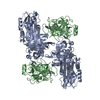
| Related structure data |  2cb4SC S: Starting model for refinement C: citing same article ( |
|---|---|
| Similar structure data |


-Links
 PDBj
PDBj


- Assembly
| Deposited unit | 
| ||||||||
|---|---|---|---|---|---|---|---|---|---|
| 1 | 
| ||||||||
| 2 | 
| ||||||||
| 3 | 
| ||||||||
| 4 | 
| ||||||||
| Unit cell |
|
-Components
| #1: Protein | Mass: 32919.254 Da / Num. of mol.: 16 / Fragment: CATALYTIC DOMAIN RESIDUES 30-308 / Mutation: YES Source method: isolated from a genetically manipulated source Source: (gene. exp.) BACILLUS SPHAERICUS (bacteria) / Strain: SSII-1 / Production host: Compound details | ENGINEERED RESIDUE IN CHAIN A, GLU 195 TO GLN ENGINEERED RESIDUE IN CHAIN B, GLU 195 TO GLN ...ENGINEERED | |
|---|
-Experimental details
-Experiment
| Experiment | Method: X-RAY DIFFRACTION / Number of used crystals: 1 |
|---|
- Sample preparation
| Crystal | Density Matthews: 3.59 Å3/Da / Density % sol: 65.45 % |
|---|---|
| Crystal grow | pH: 6.3 Details: 100 MM NA-K PHOSPHATE (PH 6.3), 8-14% (W/V) PEG 8000, 50-150 MM NACL |
-Data collection
| Diffraction | Mean temperature: 100 K |
|---|---|
| Diffraction source | Source: SYNCHROTRON / Site: SLS  / Beamline: X06SA / Wavelength: 0.9787 / Beamline: X06SA / Wavelength: 0.9787 |
| Detector | Type: MARRESEARCH / Detector: CCD |
| Radiation | Monochromator: SI(111) / Protocol: SINGLE WAVELENGTH / Monochromatic (M) / Laue (L): M / Scattering type: x-ray |
| Radiation wavelength | Wavelength: 0.9787 Å / Relative weight: 1 |
| Reflection | Resolution: 3→50 Å / Num. obs: 126014 / % possible obs: 99.6 % / Redundancy: 5.7 % / Biso Wilson estimate: 53 Å2 / Rmerge(I) obs: 0.12 / Net I/σ(I): 13 |
| Reflection shell | Resolution: 3→3.05 Å / Redundancy: 5.4 % / Rmerge(I) obs: 0.48 / Mean I/σ(I) obs: 3.5 / % possible all: 98.3 |
- Processing
| Software |
| |||||||||||||||||||||||||||||||||||||||||||||||||||||||||||||||||||||||||||||||||||||||||||||||
|---|---|---|---|---|---|---|---|---|---|---|---|---|---|---|---|---|---|---|---|---|---|---|---|---|---|---|---|---|---|---|---|---|---|---|---|---|---|---|---|---|---|---|---|---|---|---|---|---|---|---|---|---|---|---|---|---|---|---|---|---|---|---|---|---|---|---|---|---|---|---|---|---|---|---|---|---|---|---|---|---|---|---|---|---|---|---|---|---|---|---|---|---|---|---|---|---|
| Refinement | Method to determine structure: MOLECULAR REPLACEMENT Starting model: PDB ENTRY 2CB4 Resolution: 3→50 Å / Isotropic thermal model: TNT BCORREL V1.0 / Cross valid method: THROUGHOUT / σ(F): 0 / Stereochemistry target values: TNT CSDX_PROTGEO
| |||||||||||||||||||||||||||||||||||||||||||||||||||||||||||||||||||||||||||||||||||||||||||||||
| Solvent computation | Solvent model: BABINET MODEL WITH MASK / Bsol: 187 Å2 / ksol: 1 e/Å3 | |||||||||||||||||||||||||||||||||||||||||||||||||||||||||||||||||||||||||||||||||||||||||||||||
| Refinement step | Cycle: LAST / Resolution: 3→50 Å
| |||||||||||||||||||||||||||||||||||||||||||||||||||||||||||||||||||||||||||||||||||||||||||||||
| Refine LS restraints |
|
