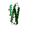
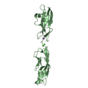
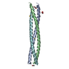
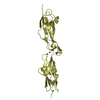
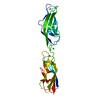
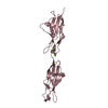
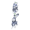
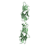
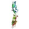
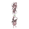
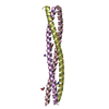
Entry Database : PDB / ID : 2c5jTitle N-terminal domain of tlg1, domain-swapped dimer T-SNARE AFFECTING A LATE GOLGI COMPARTMENT PROTEIN 1 Keywords / / / Function / homology Function Domain/homology Component
/ / / / / / / / / / / / / / / / / / / / / / / / / / / / / / / / / / / / / / / / / / / / / / / / Biological species SACCHAROMYCES CEREVISIAE (brewer's yeast)Method / / Resolution : 2.1 Å Authors Fridmann-Sirkis, Y. / Kent, H.M. / Lewis, M.J. / Evans, P.R. / Pelham, H.R.B. Journal : Traffic / Year : 2006Title : Structural Analysis of the Interaction between the Snare Tlg1 and Vps51.Authors : Fridmann-Sirkis, Y. / Kent, H.M. / Lewis, M.J. / Evans, P.R. / Pelham, H.R.B. History Deposition Oct 27, 2005 Deposition site / Processing site Revision 1.0 Jan 25, 2006 Provider / Type Revision 1.1 May 8, 2011 Group Revision 1.2 Jul 13, 2011 Group Revision 1.3 Dec 13, 2023 Group Data collection / Database references ... Data collection / Database references / Other / Refinement description Category chem_comp_atom / chem_comp_bond ... chem_comp_atom / chem_comp_bond / database_2 / pdbx_database_status / pdbx_initial_refinement_model Item / _database_2.pdbx_database_accession / _pdbx_database_status.status_code_sf
Show all Show less
 Movie
Movie Controller


 Open data
 Basic information
 Components
 Keywords
 Function and homology information
 X-RAY DIFFRACTION /
  Authors
 Citation
 Structure visualization
 Downloads & links
 Other downloads
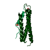


 PDBj
PDBj
 Assembly
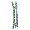

 Mass: 18.015 Da / Num. of mol.: 75 / Source method: isolated from a natural source / Formula: H2O
 Sample preparation
 Processing