 Movie
Movie Controller

[English] 日本語
 Yorodumi
- PDB-2c31: CRYSTAL STRUCTURE OF OXALYL-COA DECARBOXYLASE IN COMPLEX WITH THE... -
+ Open data

- Basic information
| Entry | Database: PDB / ID: 2c31 | ||||||
|---|---|---|---|---|---|---|---|
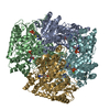
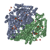
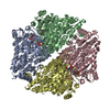
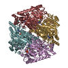
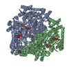
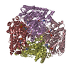
| Title | CRYSTAL STRUCTURE OF OXALYL-COA DECARBOXYLASE IN COMPLEX WITH THE COFACTOR DERIVATIVE THIAMIN-2-THIAZOLONE DIPHOSPHATE AND ADENOSINE DIPHOSPHATE | ||||||
 Components Components | OXALYL-COA DECARBOXYLASE | ||||||
 Keywords Keywords | LYASE / OXALYL-COA DECARBOXYLASE / OXALATE / THIAMIN DIPHOSPHATE / FLAVOPROTEIN / THIAMINE PYROPHOSPHATE | ||||||
| Function / homology |  Function and homology information Function and homology informationoxalyl-CoA decarboxylase / oxalyl-CoA decarboxylase activity / oxalate catabolic process / fatty acid alpha-oxidation / thiamine pyrophosphate binding / ADP binding / magnesium ion binding / identical protein binding Similarity search - Function | ||||||
| Biological species |  OXALOBACTER FORMIGENES (bacteria) OXALOBACTER FORMIGENES (bacteria) | ||||||
| Method |  X-RAY DIFFRACTION / SYNCHROTRON / MOLECULAR REPLACEMENT / Resolution: 1.73 Å X-RAY DIFFRACTION / SYNCHROTRON / MOLECULAR REPLACEMENT / Resolution: 1.73 Å | ||||||
 Authors Authors | Berthold, C.L. / Moussatche, P. / Richards, N.G.J. / Lindqvist, Y. | ||||||
 Citation Citation | Journal: J.Biol.Chem. / Year: 2005 Title: Structural Basis for Activation of the Thiamin Diphosphate-Dependent Enzyme Oxalyl-Coa Decarboxylase by Adenosine Diphosphate. Authors: Berthold, C.L. / Moussatche, P. / Richards, N.G.J. / Lindqvist, Y. #1: Journal: Biochim.Biophys.Acta / Year: 2006 Title: Detection and Characterization of Merohedral Twinning in Crystals of Oxalyl-Coenzyme a Decarboxylase from Oxalobacter Formigenes. Authors: Berthold, C.L. / Sidhu, H. / Ricagno, S. / Richards, N.G.J. / Lindqvist, Y. | ||||||
| History |
|
- Structure visualization
| Structure viewer | Molecule: MolmilJmol/JSmol |
|---|
- Downloads & links
-Download
| PDBx/mmCIF format | 2c31.cif.gz | 231.8 KB | Display | PDBx/mmCIF format |
|---|---|---|---|---|
| PDB format | pdb2c31.ent.gz | 184.4 KB | Display | PDB format |
| PDBx/mmJSON format | 2c31.json.gz | Tree view | PDBx/mmJSON format | |
| Others |  Other downloads Other downloads |
-Validation report
| Arichive directory | https://data.pdbj.org/pub/pdb/validation_reports/c3/2c31ftp://data.pdbj.org/pub/pdb/validation_reports/c3/2c31 | HTTPS FTP |
|---|
-Related structure data
| Related structure data |  1jscS S: Starting model for refinement |
|---|---|
| Similar structure data |


-Links
 PDBj
PDBj


- Assembly
| Deposited unit | 
| ||||||||
|---|---|---|---|---|---|---|---|---|---|
| 1 | 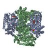
| ||||||||
| Unit cell |
| ||||||||
| Noncrystallographic symmetry (NCS) | NCS oper: (Code: given Matrix: (-1, -4.0E-5, 0.00211), Vector: |
-Components
| #1: Protein | Mass: 60749.867 Da / Num. of mol.: 2 Source method: isolated from a genetically manipulated source Source: (gene. exp.) OXALOBACTER FORMIGENES (bacteria) / Production host: #2: Chemical |   Mass: 24.305 Da / Num. of mol.: 2 / Source method: obtained synthetically / Formula: Mg Mass: 24.305 Da / Num. of mol.: 2 / Source method: obtained synthetically / Formula: Mg#3: Chemical |   Mass: 440.306 Da / Num. of mol.: 2 / Source method: obtained synthetically / Formula: C12H18N4O8P2S Mass: 440.306 Da / Num. of mol.: 2 / Source method: obtained synthetically / Formula: C12H18N4O8P2S#4: Chemical |   Mass: 427.201 Da / Num. of mol.: 2 / Source method: obtained synthetically / Formula: C10H15N5O10P2 / Comment: ADP, energy-carrying molecule*YM Mass: 427.201 Da / Num. of mol.: 2 / Source method: obtained synthetically / Formula: C10H15N5O10P2 / Comment: ADP, energy-carrying molecule*YM#5: Water | ChemComp-HOH / |  Mass: 18.015 Da / Num. of mol.: 658 / Source method: isolated from a natural source / Formula: H2O Mass: 18.015 Da / Num. of mol.: 658 / Source method: isolated from a natural source / Formula: H2O |
|---|
-Experimental details
-Experiment
| Experiment | Method: X-RAY DIFFRACTION / Number of used crystals: 1 |
|---|
- Sample preparation
| Crystal | Density Matthews: 2.8 Å3/Da / Density % sol: 53.61 % Description: ACETOHYDROXYACID SYNTHASE WAS USED AS TEMPLATE TO SOLVE AN APPROXIMATE INCOMPLETE NOT FULLY REFINED STRUCTURE OF OXALYL-COA DECARBOXYLASE IN A DIFFERENT SPACE GROUP TO 4.1 A RESOLUTION. ...Description: ACETOHYDROXYACID SYNTHASE WAS USED AS TEMPLATE TO SOLVE AN APPROXIMATE INCOMPLETE NOT FULLY REFINED STRUCTURE OF OXALYL-COA DECARBOXYLASE IN A DIFFERENT SPACE GROUP TO 4.1 A RESOLUTION. THIS LOW RESOLUTION MODEL WAS THEN USED FOR MOLECULAR REPLACE IN THIS DATA. |
|---|---|
| Crystal grow | pH: 6.5 Details: 27V/V % PEG 550MME, 100MM BIS-TRIS PH 6.5, 50MM CACL2 |
-Data collection
| Diffraction | Mean temperature: 100 K |
|---|---|
| Diffraction source | Source: SYNCHROTRON / Site: ESRF  / Beamline: ID23-1 / Wavelength: 0.97565 / Beamline: ID23-1 / Wavelength: 0.97565 |
| Detector | Type: MARRESEARCH / Detector: CCD / Date: Jul 14, 2004 / Details: SILICON TOROIDAL MIRROR COATED WITH RHODIUM |
| Radiation | Monochromator: SILICON (1 1 1) CHANNEL- CUT / Protocol: SINGLE WAVELENGTH / Monochromatic (M) / Laue (L): M / Scattering type: x-ray |
| Radiation wavelength | Wavelength: 0.97565 Å / Relative weight: 1 |
| Reflection | Resolution: 1.73→29.24 Å / Num. obs: 140751 / % possible obs: 99.8 % / Observed criterion σ(I): 2 / Redundancy: 7.1 % / Biso Wilson estimate: 21.13 Å2 / Rmerge(I) obs: 0.1 / Net I/σ(I): 17.9 |
| Reflection shell | Resolution: 1.73→1.82 Å / Redundancy: 7.1 % / Rmerge(I) obs: 0.49 / Mean I/σ(I) obs: 3.8 / % possible all: 100 |
- Processing
| Software |
| ||||||||||||||||||||||||||||||||||||||||||||||||||||||||||||||||||||||||||||||||
|---|---|---|---|---|---|---|---|---|---|---|---|---|---|---|---|---|---|---|---|---|---|---|---|---|---|---|---|---|---|---|---|---|---|---|---|---|---|---|---|---|---|---|---|---|---|---|---|---|---|---|---|---|---|---|---|---|---|---|---|---|---|---|---|---|---|---|---|---|---|---|---|---|---|---|---|---|---|---|---|---|---|
| Refinement | Method to determine structure: MOLECULAR REPLACEMENT Starting model: PDB ENTRY 1JSC Resolution: 1.73→29.2 Å / Isotropic thermal model: RESTRAINED / Cross valid method: THROUGHOUT / σ(F): 0 Stereochemistry target values: LEAST SQUARES RESIDUAL FOR HEMIHEDRAL TWINNING Details: THE PROTOCOLL FOR HEMIHEDRALLY TWINNED DATA WAS USED. THE R-VALUES SHOWN IN REMARK 3 ABOVE ARE FOR TWINNED DATA.
| ||||||||||||||||||||||||||||||||||||||||||||||||||||||||||||||||||||||||||||||||
| Solvent computation | Solvent model: CNS BULK SOLVENT MODEL / Bsol: 56.1554 Å2 / ksol: 0.357619 e/Å3 | ||||||||||||||||||||||||||||||||||||||||||||||||||||||||||||||||||||||||||||||||
| Displacement parameters | Biso mean: 25.19 Å2
| ||||||||||||||||||||||||||||||||||||||||||||||||||||||||||||||||||||||||||||||||
| Refine analyze |
| ||||||||||||||||||||||||||||||||||||||||||||||||||||||||||||||||||||||||||||||||
| Refinement step | Cycle: LAST / Resolution: 1.73→29.2 Å
| ||||||||||||||||||||||||||||||||||||||||||||||||||||||||||||||||||||||||||||||||
| Refine LS restraints |
| ||||||||||||||||||||||||||||||||||||||||||||||||||||||||||||||||||||||||||||||||
| LS refinement shell | Resolution: 1.73→1.79 Å / Total num. of bins used: 10 /
|
