 Movie
Movie Controller

[English] 日本語
 Yorodumi
- PDB-1jsc: Crystal Structure of the Catalytic Subunit of Yeast Acetohydroxya... -
+ Open data

- Basic information
| Entry | Database: PDB / ID: 1jsc | ||||||
|---|---|---|---|---|---|---|---|
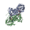
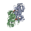
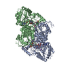
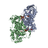
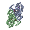
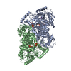
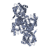
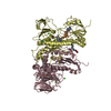
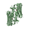
| Title | Crystal Structure of the Catalytic Subunit of Yeast Acetohydroxyacid Synthase: A target for Herbicidal Inhibitors | ||||||
 Components Components | ACETOHYDROXY-ACID SYNTHASE | ||||||
 Keywords Keywords | LYASE / ACETOHYDROXYACID SYNTHASE / ACETOLACTATE SYNTHASE / FAD / THIAMIN DIPHOSPHATE / HERBICIDE INHIBITION | ||||||
| Function / homology |  Function and homology information Function and homology informationacetolactate synthase / acetolactate synthase complex / branched-chain amino acid biosynthetic process / acetolactate synthase activity / L-valine biosynthetic process / : / thiamine pyrophosphate binding / flavin adenine dinucleotide binding / magnesium ion binding / mitochondrion Similarity search - Function | ||||||
| Biological species |  | ||||||
| Method |  X-RAY DIFFRACTION / SYNCHROTRON / MOLECULAR REPLACEMENT / Resolution: 2.6 Å X-RAY DIFFRACTION / SYNCHROTRON / MOLECULAR REPLACEMENT / Resolution: 2.6 Å | ||||||
 Authors Authors | Pang, S.S. / Duggleby, R.G. / Guddat, L.W. | ||||||
 Citation Citation | Journal: J.Mol.Biol. / Year: 2002 Title: Crystal structure of yeast acetohydroxyacid synthase: a target for herbicidal inhibitors. Authors: Pang, S.S. / Duggleby, R.G. / Guddat, L.W. | ||||||
| History |
|
- Structure visualization
| Structure viewer | Molecule: MolmilJmol/JSmol |
|---|
- Downloads & links
-Download
| PDBx/mmCIF format | 1jsc.cif.gz | 232.8 KB | Display | PDBx/mmCIF format |
|---|---|---|---|---|
| PDB format | pdb1jsc.ent.gz | 181.3 KB | Display | PDB format |
| PDBx/mmJSON format | 1jsc.json.gz | Tree view | PDBx/mmJSON format | |
| Others |  Other downloads Other downloads |
-Validation report
| Arichive directory | https://data.pdbj.org/pub/pdb/validation_reports/js/1jscftp://data.pdbj.org/pub/pdb/validation_reports/js/1jsc | HTTPS FTP |
|---|
-Related structure data
| Related structure data | 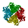 1bfdS S: Starting model for refinement |
|---|---|
| Similar structure data |

-Links
 PDBj
PDBj


- Assembly
| Deposited unit | 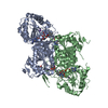
| ||||||||
|---|---|---|---|---|---|---|---|---|---|
| 1 |
| ||||||||
| Unit cell |
|
-Components
-Protein , 1 types, 2 molecules AB
| #1: Protein | Mass: 68453.000 Da / Num. of mol.: 2 / Fragment: MATURE CATALYTIC SUBUNIT Source method: isolated from a genetically manipulated source Source: (gene. exp.) Gene: ILV2 / Plasmid: pET30C / Species (production host): Escherichia coli / Production host:  |
|---|
-Non-polymers , 6 types, 336 molecules 


| #2: Chemical |  Mass: 39.098 Da / Num. of mol.: 2 / Source method: obtained synthetically / Formula: K Mass: 39.098 Da / Num. of mol.: 2 / Source method: obtained synthetically / Formula: K#3: Chemical |  Mass: 24.305 Da / Num. of mol.: 2 / Source method: obtained synthetically / Formula: Mg Mass: 24.305 Da / Num. of mol.: 2 / Source method: obtained synthetically / Formula: Mg#4: Chemical |  Mass: 96.987 Da / Num. of mol.: 3 / Source method: obtained synthetically / Formula: H2O4P Mass: 96.987 Da / Num. of mol.: 3 / Source method: obtained synthetically / Formula: H2O4P#5: Chemical |  Mass: 425.314 Da / Num. of mol.: 2 / Source method: obtained synthetically / Formula: C12H19N4O7P2S Mass: 425.314 Da / Num. of mol.: 2 / Source method: obtained synthetically / Formula: C12H19N4O7P2S#6: Chemical |  Mass: 785.550 Da / Num. of mol.: 2 / Source method: obtained synthetically / Formula: C27H33N9O15P2 / Comment: FAD*YM Mass: 785.550 Da / Num. of mol.: 2 / Source method: obtained synthetically / Formula: C27H33N9O15P2 / Comment: FAD*YM#7: Water | ChemComp-HOH / | Mass: 18.015 Da / Num. of mol.: 325 / Source method: isolated from a natural source / Formula: H2O |
|---|
-Experimental details
-Experiment
| Experiment | Method: X-RAY DIFFRACTION / Number of used crystals: 1 |
|---|
- Sample preparation
| Crystal | Density Matthews: 3.62 Å3/Da / Density % sol: 66.1 % | ||||||||||||||||||||||||||||||||||||||||||||||||||||||||
|---|---|---|---|---|---|---|---|---|---|---|---|---|---|---|---|---|---|---|---|---|---|---|---|---|---|---|---|---|---|---|---|---|---|---|---|---|---|---|---|---|---|---|---|---|---|---|---|---|---|---|---|---|---|---|---|---|---|
| Crystal grow | Temperature: 291 K / Method: vapor diffusion, hanging drop / pH: 5.8 Details: PEG 4000, potassium phosphate, thiamin diphosphate, FAD, magnesium chloride, DTT, ammonium acetate, pH 5.8, VAPOR DIFFUSION, HANGING DROP, temperature 291K | ||||||||||||||||||||||||||||||||||||||||||||||||||||||||
| Crystal grow | *PLUS Temperature: 293 K / Details: Pang, S.S., (2001) Acta Crystallogr, D57, 1321. | ||||||||||||||||||||||||||||||||||||||||||||||||||||||||
| Components of the solutions | *PLUS
|
-Data collection
| Diffraction | Mean temperature: 100 K |
|---|---|
| Diffraction source | Source: SYNCHROTRON / Site: APS  / Beamline: 14-BM-C / Wavelength: 1 Å / Beamline: 14-BM-C / Wavelength: 1 Å |
| Detector | Type: ADSC QUANTUM 4 / Detector: CCD / Date: Dec 17, 2000 / Details: mirrors |
| Radiation | Monochromator: Ge(III) / Protocol: SINGLE WAVELENGTH / Monochromatic (M) / Laue (L): M / Scattering type: x-ray |
| Radiation wavelength | Wavelength: 1 Å / Relative weight: 1 |
| Reflection | Resolution: 2.6→50 Å / Num. all: 214515 / Num. obs: 214515 / % possible obs: 97.8 % / Observed criterion σ(F): 0 / Observed criterion σ(I): 0 / Redundancy: 3.7 % / Rmerge(I) obs: 0.058 / Net I/σ(I): 17.1 |
| Reflection shell | Resolution: 2.6→2.69 Å / Redundancy: 3.6 % / Rmerge(I) obs: 0.301 / Mean I/σ(I) obs: 3.9 / Num. unique all: 5733 / % possible all: 99.2 |
| Reflection | *PLUS Num. obs: 57490 / Num. measured all: 214515 / Rmerge(I) obs: 0.058 |
| Reflection shell | *PLUS % possible obs: 99.2 % / Num. unique obs: 5733 / Num. measured obs: 20596 / Rmerge(I) obs: 0.301 |
- Processing
| Software |
| |||||||||||||||||||||||||
|---|---|---|---|---|---|---|---|---|---|---|---|---|---|---|---|---|---|---|---|---|---|---|---|---|---|---|
| Refinement | Method to determine structure: MOLECULAR REPLACEMENT Starting model: PDB ENTRY 1BFD Resolution: 2.6→100 Å / Isotropic thermal model: anisotropic / Cross valid method: R free throughout / σ(F): 0 / σ(I): 0 / Stereochemistry target values: Engh & Huber
| |||||||||||||||||||||||||
| Solvent computation | Solvent model: Jiang et al. / Bsol: -1 Å2 / ksol: -1 e/Å3 | |||||||||||||||||||||||||
| Displacement parameters |
| |||||||||||||||||||||||||
| Refine analyze |
| |||||||||||||||||||||||||
| Refinement step | Cycle: LAST / Resolution: 2.6→100 Å
| |||||||||||||||||||||||||
| Refine LS restraints |
| |||||||||||||||||||||||||
| LS refinement shell | Resolution: 2.6→2.7 Å / Rfactor Rfree error: 0
| |||||||||||||||||||||||||
| Refinement | *PLUS Highest resolution: 2.6 Å / Lowest resolution: 50 Å / Rfactor all: 0.191 / Rfactor obs: 0.188 / Rfactor Rfree: 0.219 / Rfactor Rwork: 0.188 | |||||||||||||||||||||||||
| Solvent computation | *PLUS | |||||||||||||||||||||||||
| Displacement parameters | *PLUS | |||||||||||||||||||||||||
| Refine LS restraints | *PLUS
| |||||||||||||||||||||||||
| LS refinement shell | *PLUS Highest resolution: 2.6 Å / Lowest resolution: 2.69 Å / Rfactor Rfree: 0.267 / Rfactor Rwork: 0.247 |
