 Movie
Movie Controller

[English] 日本語
 Yorodumi
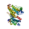
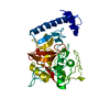
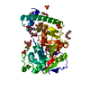
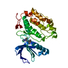
- PDB-2c0y: THE CRYSTAL STRUCTURE OF A CYS25ALA MUTANT OF HUMAN PROCATHEPSIN S -
+ Open data

- Basic information
| Entry | Database: PDB / ID: 2c0y | ||||||
|---|---|---|---|---|---|---|---|
| Title | THE CRYSTAL STRUCTURE OF A CYS25ALA MUTANT OF HUMAN PROCATHEPSIN S | ||||||
 Components Components | PROCATHEPSIN S | ||||||
 Keywords Keywords | HYDROLASE / PROCATHEPSIN S / PROENZYME / PROTEINASE / THIOL PROTEASE / PROSEGMENT BINDING LOOP / GLYCOPROTEIN / LYSOSOME / PROTEASE / ZYMOGEN | ||||||
| Function / homology |  Function and homology information Function and homology informationcathepsin S / regulation of antigen processing and presentation / positive regulation of cation channel activity / basement membrane disassembly / antigen processing and presentation of peptide antigen / endolysosome lumen / cellular response to thyroid hormone stimulus / Trafficking and processing of endosomal TLR / proteoglycan binding / Assembly of collagen fibrils and other multimeric structures ...cathepsin S / regulation of antigen processing and presentation / positive regulation of cation channel activity / basement membrane disassembly / antigen processing and presentation of peptide antigen / endolysosome lumen / cellular response to thyroid hormone stimulus / Trafficking and processing of endosomal TLR / proteoglycan binding / Assembly of collagen fibrils and other multimeric structures / response to acidic pH / toll-like receptor signaling pathway / antigen processing and presentation / collagen catabolic process / fibronectin binding / extracellular matrix disassembly / collagen binding / Degradation of the extracellular matrix / phagocytic vesicle / laminin binding / cysteine-type peptidase activity / MHC class II antigen presentation / lysosomal lumen / : / Endosomal/Vacuolar pathway / protein processing / Degradation of CDH1 / antigen processing and presentation of exogenous peptide antigen via MHC class II / tertiary granule lumen / late endosome / peptidase activity / extracellular matrix / ficolin-1-rich granule lumen / adaptive immune response / lysosome / immune response / serine-type endopeptidase activity / cysteine-type endopeptidase activity / Neutrophil degranulation / proteolysis / : / extracellular region Similarity search - Function | ||||||
| Biological species |  HOMO SAPIENS (human) HOMO SAPIENS (human) | ||||||
| Method |  X-RAY DIFFRACTION / SYNCHROTRON / MOLECULAR REPLACEMENT / Resolution: 2.1 Å X-RAY DIFFRACTION / SYNCHROTRON / MOLECULAR REPLACEMENT / Resolution: 2.1 Å | ||||||
 Authors Authors | Kaulmann, G. / Palm, G.J. / Schilling, K. / Hilgenfeld, R. / Wiederanders, B. | ||||||
 Citation Citation | Journal: Protein Sci. / Year: 2006 Title: The Crystal Structure of a Cys25 -> Ala Mutant of Human Procathepsin S Elucidates Enzyme-Prosequence Interactions. Authors: Kaulmann, G. / Palm, G.J. / Schilling, K. / Hilgenfeld, R. / Wiederanders, B. #1: Journal: Protein Sci. / Year: 1998 Title: Crystal Structure of Human Cathepsin S Authors: Mcgrath, M.E. / Palmer, J.T. / Bromme, D. / Somoza, J.R. #2: Journal: Acta Crystallogr.,Sect.D / Year: 2002Title: Structure of a Cys25 to Ser Mutant of Human Cathepsin S Authors: Turkenburg, J.P. / Lamers, M.B. / Brzozowski, A.M. / Wright, L.M. / Hubbard, R.E. / Sturt, S.L. / Williams, D.H. #3: Journal: Biochemistry / Year: 2003Title: Specificity Determinants of Human Cathepsin S Revealed by Crystal Structures of Complexes Authors: Pauly, T.A. / Sulea, T. / Ammirati, M. / Sivaraman, J. / Danley, D.E. / Griffor, M.C. / Kamath, A.V. / Wang, I.K. / Laird, E.R. / Seddon, A.P. / Menard, R. / Cygler, M. / Rath, V.L. | ||||||
| History |
| ||||||
| Remark 700 | SHEET THE SHEET STRUCTURE OF THIS MOLECULE IS BIFURCATED. IN ORDER TO REPRESENT THIS FEATURE IN ... SHEET THE SHEET STRUCTURE OF THIS MOLECULE IS BIFURCATED. IN ORDER TO REPRESENT THIS FEATURE IN THE SHEET RECORDS BELOW, TWO SHEETS ARE DEFINED. |
- Structure visualization
| Structure viewer | Molecule: MolmilJmol/JSmol |
|---|
- Downloads & links
-Download
| PDBx/mmCIF format | 2c0y.cif.gz | 79.9 KB | Display | PDBx/mmCIF format |
|---|---|---|---|---|
| PDB format | pdb2c0y.ent.gz | 59.6 KB | Display | PDB format |
| PDBx/mmJSON format | 2c0y.json.gz | Tree view | PDBx/mmJSON format | |
| Others |  Other downloads Other downloads |
-Validation report
| Arichive directory | https://data.pdbj.org/pub/pdb/validation_reports/c0/2c0yftp://data.pdbj.org/pub/pdb/validation_reports/c0/2c0y | HTTPS FTP |
|---|
-Related structure data
| Related structure data |  1by8S S: Starting model for refinement |
|---|---|
| Similar structure data |


-Links
 PDBj
PDBj


- Assembly
| Deposited unit | 
| ||||||||
|---|---|---|---|---|---|---|---|---|---|
| 1 |
| ||||||||
| Unit cell |
| ||||||||
| Components on special symmetry positions |
|
-Components
| #1: Protein | Mass: 35839.477 Da / Num. of mol.: 1 / Fragment: PROENZYME, RESIDUES 17-331 / Mutation: YES Source method: isolated from a genetically manipulated source Source: (gene. exp.) HOMO SAPIENS (human) / Organ: TESTIS / Cell line (production host): High Five / Production host:  TRICHOPLUSIA NI (cabbage looper) / References: UniProt: P25774, cathepsin S TRICHOPLUSIA NI (cabbage looper) / References: UniProt: P25774, cathepsin S |
|---|---|
| #2: Water | ChemComp-HOH /  Mass: 18.015 Da / Num. of mol.: 242 / Source method: isolated from a natural source / Formula: H2O Mass: 18.015 Da / Num. of mol.: 242 / Source method: isolated from a natural source / Formula: H2O |
| Compound details | PROTEASE RESPONSIBLE FOR THE REMOVAL OF THE INVARIANT CHAIN FROM MHC CLASS II MOLECULES ENGINEERED ...PROTEASE RESPONSIBL |
| Has protein modification | Y |
| Sequence details | CLONED WITHOUT SIGNAL PEPTIDE |
-Experimental details
-Experiment
| Experiment | Method: X-RAY DIFFRACTION / Number of used crystals: 1 |
|---|
- Sample preparation
| Crystal | Density Matthews: 2.31 Å3/Da / Density % sol: 47 % |
|---|---|
| Crystal grow | Method: vapor diffusion, hanging drop / pH: 7.5 Details: CRYSTALLISATION WAS PERFORMED BY THE HANGING-DROP VAPOUR DIFFUSION METHOD. EQUAL VOLUMES PROCATHEPSIN S (CYS25ALA) AT 7.0 MG/ML AND WELL SOLUTION WERE COMBINED AND PLACED OVER A WELL ...Details: CRYSTALLISATION WAS PERFORMED BY THE HANGING-DROP VAPOUR DIFFUSION METHOD. EQUAL VOLUMES PROCATHEPSIN S (CYS25ALA) AT 7.0 MG/ML AND WELL SOLUTION WERE COMBINED AND PLACED OVER A WELL CONTAINING 0.1M TRIS/HCL, 0.2M MAGNESIUM ACETATE AND 20% PEG 8000, PH 7.5 |
-Data collection
| Diffraction | Mean temperature: 100 K |
|---|---|
| Diffraction source | Source: SYNCHROTRON / Site: EMBL/DESY, HAMBURG  / Beamline: X13 / Wavelength: 0.803 / Beamline: X13 / Wavelength: 0.803 |
| Detector | Type: MARRESEARCH / Detector: CCD / Date: Jun 18, 2001 Details: TRIANGULAR SI (111) MONOCHROMATOR AND A CONTINUOUS BENT RH-COATED MIRROR |
| Radiation | Protocol: SINGLE WAVELENGTH / Monochromatic (M) / Laue (L): M / Scattering type: x-ray |
| Radiation wavelength | Wavelength: 0.803 Å / Relative weight: 1 |
| Reflection | Resolution: 2.1→30 Å / Num. obs: 19350 / % possible obs: 97.9 % / Observed criterion σ(I): -3 / Redundancy: 6.4 % / Biso Wilson estimate: 24.7 Å2 / Rmerge(I) obs: 0.09 / Net I/σ(I): 13.4 |
| Reflection shell | Resolution: 2.1→2.14 Å / Redundancy: 4.1 % / Rmerge(I) obs: 0.47 / Mean I/σ(I) obs: 3 / % possible all: 88.5 |
- Processing
| Software |
| ||||||||||||||||||||||||||||||||||||||||||||||||||||||||||||||||||||||||||||||||
|---|---|---|---|---|---|---|---|---|---|---|---|---|---|---|---|---|---|---|---|---|---|---|---|---|---|---|---|---|---|---|---|---|---|---|---|---|---|---|---|---|---|---|---|---|---|---|---|---|---|---|---|---|---|---|---|---|---|---|---|---|---|---|---|---|---|---|---|---|---|---|---|---|---|---|---|---|---|---|---|---|---|
| Refinement | Method to determine structure: MOLECULAR REPLACEMENT Starting model: PDB ENTRY 1BY8 Resolution: 2.1→19.76 Å / Rfactor Rfree error: 0.008 / Data cutoff high absF: 1874964.9 / Isotropic thermal model: RESTRAINED / Cross valid method: THROUGHOUT / σ(F): 0
| ||||||||||||||||||||||||||||||||||||||||||||||||||||||||||||||||||||||||||||||||
| Solvent computation | Solvent model: FLAT MODEL / Bsol: 62.2678 Å2 / ksol: 0.406382 e/Å3 | ||||||||||||||||||||||||||||||||||||||||||||||||||||||||||||||||||||||||||||||||
| Displacement parameters | Biso mean: 28.7 Å2
| ||||||||||||||||||||||||||||||||||||||||||||||||||||||||||||||||||||||||||||||||
| Refine analyze |
| ||||||||||||||||||||||||||||||||||||||||||||||||||||||||||||||||||||||||||||||||
| Refinement step | Cycle: LAST / Resolution: 2.1→19.76 Å
| ||||||||||||||||||||||||||||||||||||||||||||||||||||||||||||||||||||||||||||||||
| Refine LS restraints |
| ||||||||||||||||||||||||||||||||||||||||||||||||||||||||||||||||||||||||||||||||
| LS refinement shell | Resolution: 2.1→2.14 Å / Rfactor Rfree error: 0.031 / Total num. of bins used: 10
| ||||||||||||||||||||||||||||||||||||||||||||||||||||||||||||||||||||||||||||||||
| Xplor file |
|
