 Movie
Movie Controller


+ Open data

- Basic information
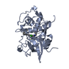
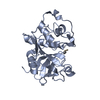
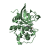
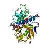
| Entry | Database: PDB / ID: 1npz | ||||||
|---|---|---|---|---|---|---|---|
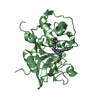
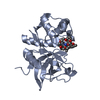
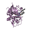
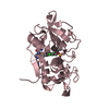
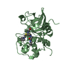
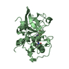
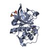
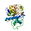
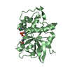
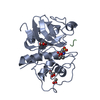
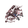
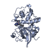
| Title | Crystal structures of Cathepsin S inhibitor complexes | ||||||
 Components Components | Cathepsin S | ||||||
 Keywords Keywords | HYDROLASE / Antigen presentation / binding specificity / cysteine proteases / inhibitor complexes / structure-based design / structural plasticity | ||||||
| Function / homology |  Function and homology information Function and homology informationcathepsin S / regulation of antigen processing and presentation / positive regulation of cation channel activity / basement membrane disassembly / antigen processing and presentation of peptide antigen / endolysosome lumen / cellular response to thyroid hormone stimulus / Trafficking and processing of endosomal TLR / proteoglycan binding / Assembly of collagen fibrils and other multimeric structures ...cathepsin S / regulation of antigen processing and presentation / positive regulation of cation channel activity / basement membrane disassembly / antigen processing and presentation of peptide antigen / endolysosome lumen / cellular response to thyroid hormone stimulus / Trafficking and processing of endosomal TLR / proteoglycan binding / Assembly of collagen fibrils and other multimeric structures / response to acidic pH / toll-like receptor signaling pathway / antigen processing and presentation / collagen catabolic process / fibronectin binding / extracellular matrix disassembly / Degradation of the extracellular matrix / collagen binding / phagocytic vesicle / laminin binding / cysteine-type peptidase activity / MHC class II antigen presentation / lysosomal lumen / : / Endosomal/Vacuolar pathway / protein processing / Degradation of CDH1 / antigen processing and presentation of exogenous peptide antigen via MHC class II / tertiary granule lumen / late endosome / peptidase activity / extracellular matrix / ficolin-1-rich granule lumen / adaptive immune response / lysosome / immune response / serine-type endopeptidase activity / cysteine-type endopeptidase activity / Neutrophil degranulation / proteolysis / : / extracellular region Similarity search - Function | ||||||
| Biological species |  Homo sapiens (human) Homo sapiens (human) | ||||||
| Method |  X-RAY DIFFRACTION / MOLECULAR REPLACEMENT / Resolution: 2 Å X-RAY DIFFRACTION / MOLECULAR REPLACEMENT / Resolution: 2 Å | ||||||
 Authors Authors | Pauly, T.A. / Sulea, T. / Ammirati, M. / Sivaraman, J. / Danley, D.E. / Griffor, M.C. / Kamath, A.V. / Wang, I.K. / Laird, E.R. / Seddon, A.P. ...Pauly, T.A. / Sulea, T. / Ammirati, M. / Sivaraman, J. / Danley, D.E. / Griffor, M.C. / Kamath, A.V. / Wang, I.K. / Laird, E.R. / Seddon, A.P. / Menard, R. / Cygler, M. / Rath, V.L. | ||||||
 Citation Citation | Journal: Biochemistry / Year: 2003 Title: Specificity determinants of human cathepsin s revealed by crystal structures of complexes. Authors: Pauly, T.A. / Sulea, T. / Ammirati, M. / Sivaraman, J. / Danley, D.E. / Griffor, M.C. / Kamath, A.V. / Wang, I.K. / Laird, E.R. / Seddon, A.P. / Menard, R. / Cygler, M. / Rath, V.L. | ||||||
| History |
|
- Structure visualization
| Structure viewer | Molecule: MolmilJmol/JSmol |
|---|
- Downloads & links
-Download
| PDBx/mmCIF format | 1npz.cif.gz | 100.6 KB | Display | PDBx/mmCIF format |
|---|---|---|---|---|
| PDB format | pdb1npz.ent.gz | 76.9 KB | Display | PDB format |
| PDBx/mmJSON format | 1npz.json.gz | Tree view | PDBx/mmJSON format | |
| Others |  Other downloads Other downloads |
-Validation report
| Arichive directory | https://data.pdbj.org/pub/pdb/validation_reports/np/1npzftp://data.pdbj.org/pub/pdb/validation_reports/np/1npz | HTTPS FTP |
|---|
-Related structure data
| Related structure data |  1nqcC 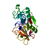 1atkS 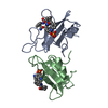 1cj1S C: citing same article ( S: Starting model for refinement |
|---|---|
| Similar structure data |


-Links
 PDBj
PDBj


- Assembly
| Deposited unit | 
| ||||||||
|---|---|---|---|---|---|---|---|---|---|
| 1 | 
| ||||||||
| 2 | 
| ||||||||
| Unit cell |
|
-Components
| #1: Protein | Mass: 24020.963 Da / Num. of mol.: 2 / Source method: isolated from a natural source / Source: (natural) Homo sapiens (human) / References: UniProt: P25774, cathepsin S#2: Chemical |   Mass: 529.691 Da / Num. of mol.: 2 / Source method: obtained synthetically / Formula: C28H39N3O5S Mass: 529.691 Da / Num. of mol.: 2 / Source method: obtained synthetically / Formula: C28H39N3O5S#3: Water | ChemComp-HOH / |  Mass: 18.015 Da / Num. of mol.: 175 / Source method: isolated from a natural source / Formula: H2O Mass: 18.015 Da / Num. of mol.: 175 / Source method: isolated from a natural source / Formula: H2OHas protein modification | Y | |
|---|
-Experimental details
-Experiment
| Experiment | Method: X-RAY DIFFRACTION / Number of used crystals: 1 |
|---|
- Sample preparation
| Crystal | Density Matthews: 2.33 Å3/Da / Density % sol: 46.9 % | ||||||||||||||||||||||||||||||||||||||||||||||||
|---|---|---|---|---|---|---|---|---|---|---|---|---|---|---|---|---|---|---|---|---|---|---|---|---|---|---|---|---|---|---|---|---|---|---|---|---|---|---|---|---|---|---|---|---|---|---|---|---|---|
| Crystal grow | Temperature: 293 K / Method: vapor diffusion, hanging drop / pH: 5.5 Details: Sodium Acetate, Ammonium Sulphate, pH 5.5, VAPOR DIFFUSION, HANGING DROP, temperature 293K | ||||||||||||||||||||||||||||||||||||||||||||||||
| Crystal grow | *PLUS Temperature: 22 ℃ / pH: 6.5 | ||||||||||||||||||||||||||||||||||||||||||||||||
| Components of the solutions | *PLUS
|
-Data collection
| Diffraction | Mean temperature: 293 K |
|---|---|
| Diffraction source | Source: ROTATING ANODE / Type: RIGAKU RU300 / Wavelength: 1.5418 Å |
| Detector | Type: MARRESEARCH / Detector: AREA DETECTOR / Date: Jul 7, 2001 |
| Radiation | Monochromator: Graphite / Protocol: SINGLE WAVELENGTH / Monochromatic (M) / Laue (L): M / Scattering type: x-ray |
| Radiation wavelength | Wavelength: 1.5418 Å / Relative weight: 1 |
| Reflection | Resolution: 2→30 Å / Num. all: 85622 / Num. obs: 83669 / % possible obs: 98.5 % / Observed criterion σ(F): 0 / Observed criterion σ(I): 0 / Redundancy: 2.8 % / Rsym value: 0.091 / Net I/σ(I): 7.5 |
| Reflection shell | Resolution: 2→2.07 Å / Rsym value: 0.503 / % possible all: 96.1 |
| Reflection | *PLUS Num. obs: 30379 / Num. measured all: 83669 / Rmerge(I) obs: 0.091 |
- Processing
| Software |
| ||||||||||||||||||||||||
|---|---|---|---|---|---|---|---|---|---|---|---|---|---|---|---|---|---|---|---|---|---|---|---|---|---|
| Refinement | Method to determine structure: MOLECULAR REPLACEMENT Starting model: Search model consists of a polyalanine homology model of cathepsin S constructed from cathepsin K (PDB code 1atk) and Cathepsin L from the procathepsin L (PDB code 1cj1) Resolution: 2→30 Å / σ(F): 0 / σ(I): 0
| ||||||||||||||||||||||||
| Refine analyze | Luzzati coordinate error obs: 0.27 Å | ||||||||||||||||||||||||
| Refinement step | Cycle: LAST / Resolution: 2→30 Å
| ||||||||||||||||||||||||
| Refine LS restraints |
| ||||||||||||||||||||||||
| Refinement | *PLUS | ||||||||||||||||||||||||
| Solvent computation | *PLUS | ||||||||||||||||||||||||
| Displacement parameters | *PLUS | ||||||||||||||||||||||||
| Refine LS restraints | *PLUS
|
