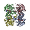
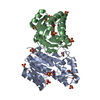
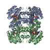
acetoacetyl-CoA reductase activity / 3-oxoacyl-[acyl-carrier-protein] reductase / 3-oxoacyl-[acyl-carrier-protein] reductase (NADPH) activity / oxidoreductase activity, acting on the CH-OH group of donors, NAD or NADP as acceptor / NADPH binding / quinone binding / NAD binding / fatty acid biosynthetic process Similarity search - Function
Mass: 18.015 Da / Num. of mol.: 184 / Source method: isolated from a natural source / Formula: H2O
Sequence details
THE AUTHORS OF THIS GENBANK ENTRY ARE ALSO AUTHORS OF THE STRUCTURAL DATA. IN THIS GENBANK ENTRY, ...THE AUTHORS OF THIS GENBANK ENTRY ARE ALSO AUTHORS OF THE STRUCTURAL DATA. IN THIS GENBANK ENTRY, RESIDUE 58 HAS BEEN ENTERED INCORRECTLY AS A HISTIDINE NOT A TYROSINE. A CHECK OF THE DNA SEQUENCING BY THESE AUTHORS SUGGESTS RESIDUE 58 IS A TYROSINE AS OBSERVED BY THE STRUCTURE. WE ARE CURRENTLY WRITING TO NCBI TO AMEND THE Q86RB1_PLAF7 AND ITS CORRESPONDING NUCLEOTIDE ENTRY.
-
Experimental details
-
Experiment
Experiment
Method: X-RAY DIFFRACTION / Number of used crystals: 1
Resolution: 1.5→20 Å / Rfactor Rfree error: 0.007 / Data cutoff high absF: 3225021.62 / Isotropic thermal model: RESTRAINED / Cross valid method: THROUGHOUT / σ(F): 0 Details: THIS ENTRY CONTAINS ATOMS WHOSE B- FACTORS HAVE BEEN REFINED BUT HAVE AN OCCUPANCY OF 0.00.
In the structure databanks used in Yorodumi, some data are registered as the other names, "COVID-19 virus" and "2019-nCoV". Here are the details of the virus and the list of structure data.
Jan 31, 2019. EMDB accession codes are about to change! (news from PDBe EMDB page)
EMDB accession codes are about to change! (news from PDBe EMDB page)
The allocation of 4 digits for EMDB accession codes will soon come to an end. Whilst these codes will remain in use, new EMDB accession codes will include an additional digit and will expand incrementally as the available range of codes is exhausted. The current 4-digit format prefixed with “EMD-” (i.e. EMD-XXXX) will advance to a 5-digit format (i.e. EMD-XXXXX), and so on. It is currently estimated that the 4-digit codes will be depleted around Spring 2019, at which point the 5-digit format will come into force.
The EM Navigator/Yorodumi systems omit the EMD- prefix.
Related info.:Q: What is EMD? / ID/Accession-code notation in Yorodumi/EM Navigator
Yorodumi is a browser for structure data from EMDB, PDB, SASBDB, etc.
This page is also the successor to EM Navigator detail page, and also detail information page/front-end page for Omokage search.
The word "yorodu" (or yorozu) is an old Japanese word meaning "ten thousand". "mi" (miru) is to see.
Related info.:EMDB / PDB / SASBDB / Comparison of 3 databanks / Yorodumi Search / Aug 31, 2016. New EM Navigator & Yorodumi / Yorodumi Papers / Jmol/JSmol / Function and homology information / Changes in new EM Navigator and Yorodumi
 Movie
Movie Controller


 Open data
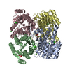
 Basic information
 Components
 Keywords
 Function and homology information
 X-RAY DIFFRACTION /
  Authors
 Citation
 Structure visualization
 Downloads & links
 Other downloads
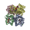


 PDBj
PDBj


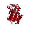
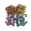
 Assembly


 Mass: 96.063 Da / Num. of mol.: 1 / Source method: obtained synthetically / Formula: SO4
 Mass: 18.015 Da / Num. of mol.: 184 / Source method: isolated from a natural source / Formula: H2O
 Sample preparation
 / Beamline: BM14 / Wavelength: 0.933
  Processing