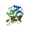
- PDB-2bzl: CRYSTAL STRUCTURE OF THE HUMAN PROTEIN TYROSINE PHOSPHATASE N14 A... -
+
Open data
ID or keywords:
Loading...
-
Basic information
Entry
Database: PDB / ID: 2bzl
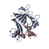
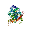
Title
CRYSTAL STRUCTURE OF THE HUMAN PROTEIN TYROSINE PHOSPHATASE N14 AT 1. 65 A RESOLUTION
Components
TYROSINE-PROTEIN PHOSPHATASE, NON-RECEPTOR TYPE 14
Keywords
HYDROLASE / PTPN14 / PROTEIN PHOSPHATASE
Function / homology
Function and homology information
lymphangiogenesis / regulation of protein export from nucleus / Interleukin-37 signaling / protein dephosphorylation / protein-tyrosine-phosphatase / protein tyrosine phosphatase activity / transcription coregulator activity / receptor tyrosine kinase binding / cytoskeleton / negative regulation of cell population proliferation ...lymphangiogenesis / regulation of protein export from nucleus / Interleukin-37 signaling / protein dephosphorylation / protein-tyrosine-phosphatase / protein tyrosine phosphatase activity / transcription coregulator activity / receptor tyrosine kinase binding / cytoskeleton / negative regulation of cell population proliferation / nucleoplasm / nucleus / cytoplasm Similarity search - Function
Protein-tyrosine phosphatase, non-receptor type-14/21 / PTPN14/21, FERM domain C-lobe / FERM, C-terminal PH-like domain / FERM C-terminal PH-like domain / FERM C-terminal PH-like domain / FERM, N-terminal / FERM N-terminal domain / FERM domain signature 1. / FERM conserved site / FERM domain signature 2. ...Protein-tyrosine phosphatase, non-receptor type-14/21 / PTPN14/21, FERM domain C-lobe / FERM, C-terminal PH-like domain / FERM C-terminal PH-like domain / FERM C-terminal PH-like domain / FERM, N-terminal / FERM N-terminal domain / FERM domain signature 1. / FERM conserved site / FERM domain signature 2. / FERM central domain / FERM/acyl-CoA-binding protein superfamily / Protein tyrosine phosphatase superfamily / FERM central domain / FERM superfamily, second domain / Protein-Tyrosine Phosphatase; Chain A / FERM domain / FERM domain profile. / Band 4.1 domain / Band 4.1 homologues / Protein tyrosine phosphatase, catalytic domain / PTP type protein phosphatase domain profile. / Protein-tyrosine phosphatase / Tyrosine-specific protein phosphatase, PTPase domain / Protein-tyrosine phosphatase, catalytic / Protein tyrosine phosphatase, catalytic domain motif / Tyrosine specific protein phosphatases active site. / Protein-tyrosine phosphatase, active site / Tyrosine specific protein phosphatases domain profile. / Tyrosine-specific protein phosphatases domain / Protein-tyrosine phosphatase-like / PH-like domain superfamily / Ubiquitin-like domain superfamily / Alpha-Beta Complex / Alpha Beta Similarity search - Domain/homology
Mass: 18.015 Da / Num. of mol.: 244 / Source method: isolated from a natural source / Formula: H2O
Compound details
BELONGS TO THE PROTEIN-TYROSINE PHOSPHATASE FAMILY. NON-RECEPTOR CLASS SUBFAMILY.
Sequence details
RESIDUES 886-1187 CORRESPOND TO THE CATALYTIC DOMAIN OF OF THE PROTEIN, RESIDUES 863-885 ARE PART ...RESIDUES 886-1187 CORRESPOND TO THE CATALYTIC DOMAIN OF OF THE PROTEIN, RESIDUES 863-885 ARE PART OF A EXPRESSION SYSTEM HIS-TAG.
-
Experimental details
-
Experiment
Experiment
Method: X-RAY DIFFRACTION / Number of used crystals: 1
-
Sample preparation
Crystal
Density Matthews: 2.4 Å3/Da / Density % sol: 49.2 %
Crystal grow
Method: vapor diffusion, sitting drop Details: SITTING DROP, 20% PEG3350, 0.2 M LI2SO4, 0.1 M BIS TRIS PROPANE PH 6.5, CRYO 20% ETHYLENE GLYCOL
Resolution: 1.65→77.15 Å / Cor.coef. Fo:Fc: 0.961 / Cor.coef. Fo:Fc free: 0.95 / SU B: 4.067 / SU ML: 0.063 / Cross valid method: THROUGHOUT / ESU R: 0.121 / ESU R Free: 0.092 / Stereochemistry target values: MAXIMUM LIKELIHOOD / Details: HYDROGENS HAVE BEEN ADDED IN THE RIDING POSITIONS.
Rfactor
Num. reflection
% reflection
Selection details
Rfree
0.217
1820
4.4 %
RANDOM
Rwork
0.185
-
-
-
obs
0.186
39553
99.5 %
-
Solvent computation
Ion probe radii: 0.8 Å / Shrinkage radii: 0.8 Å / VDW probe radii: 1.2 Å / Solvent model: BABINET MODEL WITH MASK
 Movie
Movie Controller

 Yorodumi
 Open data
 Basic information
 Components
 Keywords
 Function and homology information
 HOMO SAPIENS (human)
 X-RAY DIFFRACTION /
  Authors
 Citation
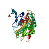
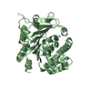
 Structure visualization
 Downloads & links
 Other downloads

 PDBj
PDBj


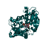
 Assembly
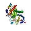


 Mass: 96.063 Da / Num. of mol.: 2 / Source method: obtained synthetically / Formula: SO4
 Mass: 62.068 Da / Num. of mol.: 2 / Source method: obtained synthetically / Formula: C2H6O2
 Mass: 18.015 Da / Num. of mol.: 244 / Source method: isolated from a natural source / Formula: H2O
 Sample preparation
 / Beamline: X10SA / Wavelength: 1.008
  Processing