 Movie
Movie Controller

[English] 日本語
 Yorodumi
- PDB-2bsl: Crystal structure of L. lactis dihydroorotate dehydrogense A in c... -
+ Open data

- Basic information
| Entry | Database: PDB / ID: 2bsl | ||||||
|---|---|---|---|---|---|---|---|
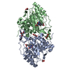
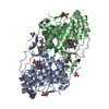
| Title | Crystal structure of L. lactis dihydroorotate dehydrogense A in complex with 3,4-dihydroxybenzoate | ||||||
 Components Components | DIHYDROOROTATE DEHYDROGENASE A | ||||||
 Keywords Keywords | OXIDOREDUCTASE / NUCLEOTIDE METABOLISM / DIHYDROOROTATE DEHYDROGENASE / FMN / FLAVOPROTEIN / PYRIMIDINE BIOSYNTHESIS | ||||||
| Function / homology |  Function and homology information Function and homology informationdihydroorotate dehydrogenase (fumarate) / dihydroorotate dehydrogenase (fumarate) activity / 'de novo' UMP biosynthetic process / 'de novo' pyrimidine nucleobase biosynthetic process / cytoplasm Similarity search - Function | ||||||
| Biological species |  LACTOCOCCUS LACTIS (lactic acid bacteria) LACTOCOCCUS LACTIS (lactic acid bacteria) | ||||||
| Method |  X-RAY DIFFRACTION / MOLECULAR REPLACEMENT / Resolution: 2.3 Å X-RAY DIFFRACTION / MOLECULAR REPLACEMENT / Resolution: 2.3 Å | ||||||
 Authors Authors | Wolfe, A.E. / Hansen, M. / Gattis, S.G. / Hu, Y.-C. / Johansson, E. / Arent, S. / Larsen, S. / Palfey, B.A. | ||||||
 Citation Citation | Journal: Biochemistry / Year: 2007 Title: Interaction of Benzoate Pyrimidine Analogues with Class 1A Dihydroorotate Dehydrogenase from Lactococcus Lactis. Authors: Wolfe, A.E. / Thymark, M. / Gattis, S.G. / Fagan, R.L. / Hu, Y.-C. / Johansson, E. / Arent, S. / Larsen, S. / Palfey, B.A. | ||||||
| History |
|
- Structure visualization
| Structure viewer | Molecule: MolmilJmol/JSmol |
|---|
- Downloads & links
-Download
| PDBx/mmCIF format | 2bsl.cif.gz | 138 KB | Display | PDBx/mmCIF format |
|---|---|---|---|---|
| PDB format | pdb2bsl.ent.gz | 106.5 KB | Display | PDB format |
| PDBx/mmJSON format | 2bsl.json.gz | Tree view | PDBx/mmJSON format | |
| Others |  Other downloads Other downloads |
-Validation report
| Arichive directory | https://data.pdbj.org/pub/pdb/validation_reports/bs/2bslftp://data.pdbj.org/pub/pdb/validation_reports/bs/2bsl | HTTPS FTP |
|---|
-Related structure data
| Related structure data |  2bx7C  1ovdS S: Starting model for refinement C: citing same article ( |
|---|---|
| Similar structure data |


-Links
 PDBj
PDBj- Assembly
| Deposited unit | 
| |||||||||||||||||||||||||||||||||||||||
|---|---|---|---|---|---|---|---|---|---|---|---|---|---|---|---|---|---|---|---|---|---|---|---|---|---|---|---|---|---|---|---|---|---|---|---|---|---|---|---|---|
| 1 |
| |||||||||||||||||||||||||||||||||||||||
| Unit cell |
| |||||||||||||||||||||||||||||||||||||||
| Noncrystallographic symmetry (NCS) | NCS domain:
NCS domain segments:
NCS oper: (Code: given Matrix: (0.87874, 0.009773, 0.47719), Vector: |
-Components
-Protein , 1 types, 2 molecules AB
| #1: Protein | Mass: 34242.168 Da / Num. of mol.: 2 Source method: isolated from a genetically manipulated source Source: (gene. exp.) LACTOCOCCUS LACTIS (lactic acid bacteria)Production host: References: UniProt: P54321, UniProt: A2RJT9*PLUS, EC: 1.3.99.11 |
|---|
-Non-polymers , 6 types, 172 molecules 


| #2: Chemical |  Mass: 456.344 Da / Num. of mol.: 2 / Source method: obtained synthetically / Formula: C17H21N4O9P Mass: 456.344 Da / Num. of mol.: 2 / Source method: obtained synthetically / Formula: C17H21N4O9P#3: Chemical | ChemComp-ACT / |  Mass: 59.044 Da / Num. of mol.: 1 / Source method: obtained synthetically / Formula: C2H3O2 Mass: 59.044 Da / Num. of mol.: 1 / Source method: obtained synthetically / Formula: C2H3O2#4: Chemical |  Mass: 24.305 Da / Num. of mol.: 2 / Source method: obtained synthetically / Formula: Mg Mass: 24.305 Da / Num. of mol.: 2 / Source method: obtained synthetically / Formula: Mg#5: Chemical | ChemComp-GOL / |  Mass: 92.094 Da / Num. of mol.: 1 / Source method: obtained synthetically / Formula: C3H8O3 Mass: 92.094 Da / Num. of mol.: 1 / Source method: obtained synthetically / Formula: C3H8O3#6: Chemical | ChemComp-DHB / |  Mass: 154.120 Da / Num. of mol.: 1 / Source method: obtained synthetically / Formula: C7H6O4 Mass: 154.120 Da / Num. of mol.: 1 / Source method: obtained synthetically / Formula: C7H6O4#7: Water | ChemComp-HOH / | Mass: 18.015 Da / Num. of mol.: 165 / Source method: isolated from a natural source / Formula: H2O |
|---|
-Details
| Compound details | CATALYSIS: (S)-DIHYDROORO |
|---|
-Experimental details
-Experiment
| Experiment | Method: X-RAY DIFFRACTION / Number of used crystals: 1 |
|---|
- Sample preparation
| Crystal | Density Matthews: 2.62 Å3/Da / Density % sol: 54 % |
|---|---|
| Crystal grow | Method: vapor diffusion, hanging drop / pH: 8.5 Details: 17 MG/ML PROTEIN AND 2.6 MM 3, 4-DIOHB HANGING DROPS 2 MICROLITERS OF PROTEIN AND 2 MICROLITERS RESERVOIR: 30 % PEG 6000, 1 MM DTT, 0.2 M SODIUM ACETATE AND 0.1 M TRIS-HCL PH 8.5 |
-Data collection
| Diffraction | Mean temperature: 120 K |
|---|---|
| Diffraction source | Source: ROTATING ANODE / Type: RIGAKU RU300 / Wavelength: 1.54179 |
| Detector | Type: MARRESEEARCH / Detector: IMAGE PLATE / Details: MIRRORS |
| Radiation | Monochromator: OSMIC MIRRORS / Protocol: SINGLE WAVELENGTH / Monochromatic (M) / Laue (L): M / Scattering type: x-ray |
| Radiation wavelength | Wavelength: 1.54179 Å / Relative weight: 1 |
| Reflection | Resolution: 2.3→25 Å / Num. obs: 32557 / % possible obs: 90.6 % / Observed criterion σ(I): -3 / Redundancy: 5.7 % / Biso Wilson estimate: 26 Å2 / Rmerge(I) obs: 0.06 / Net I/σ(I): 22.9 |
| Reflection shell | Resolution: 2.3→2.32 Å / Rmerge(I) obs: 0.23 / Mean I/σ(I) obs: 5.6 / % possible all: 62.8 |
- Processing
| Software |
| ||||||||||||||||||||||||||||||||||||||||||||||||||||||||||||||||||||||||||||||||||||||||||||||||||||||||||||||||||||||||||||||||||||||||||||||||||||||||||||||||||||||||||||||||||||||
|---|---|---|---|---|---|---|---|---|---|---|---|---|---|---|---|---|---|---|---|---|---|---|---|---|---|---|---|---|---|---|---|---|---|---|---|---|---|---|---|---|---|---|---|---|---|---|---|---|---|---|---|---|---|---|---|---|---|---|---|---|---|---|---|---|---|---|---|---|---|---|---|---|---|---|---|---|---|---|---|---|---|---|---|---|---|---|---|---|---|---|---|---|---|---|---|---|---|---|---|---|---|---|---|---|---|---|---|---|---|---|---|---|---|---|---|---|---|---|---|---|---|---|---|---|---|---|---|---|---|---|---|---|---|---|---|---|---|---|---|---|---|---|---|---|---|---|---|---|---|---|---|---|---|---|---|---|---|---|---|---|---|---|---|---|---|---|---|---|---|---|---|---|---|---|---|---|---|---|---|---|---|---|---|
| Refinement | Method to determine structure: MOLECULAR REPLACEMENT Starting model: PDB ENTRY 1OVD Resolution: 2.3→25 Å / Cor.coef. Fo:Fc: 0.954 / Cor.coef. Fo:Fc free: 0.923 / SU B: 6.805 / SU ML: 0.164 / Cross valid method: THROUGHOUT / ESU R: 0.366 / ESU R Free: 0.248 / Stereochemistry target values: MAXIMUM LIKELIHOOD / Details: HYDROGENS HAVE BEEN ADDED IN THE RIDING POSITIONS.
| ||||||||||||||||||||||||||||||||||||||||||||||||||||||||||||||||||||||||||||||||||||||||||||||||||||||||||||||||||||||||||||||||||||||||||||||||||||||||||||||||||||||||||||||||||||||
| Solvent computation | Ion probe radii: 0.8 Å / Shrinkage radii: 0.8 Å / VDW probe radii: 1.4 Å / Solvent model: MASK | ||||||||||||||||||||||||||||||||||||||||||||||||||||||||||||||||||||||||||||||||||||||||||||||||||||||||||||||||||||||||||||||||||||||||||||||||||||||||||||||||||||||||||||||||||||||
| Displacement parameters | Biso mean: 36.13 Å2
| ||||||||||||||||||||||||||||||||||||||||||||||||||||||||||||||||||||||||||||||||||||||||||||||||||||||||||||||||||||||||||||||||||||||||||||||||||||||||||||||||||||||||||||||||||||||
| Refinement step | Cycle: LAST / Resolution: 2.3→25 Å
| ||||||||||||||||||||||||||||||||||||||||||||||||||||||||||||||||||||||||||||||||||||||||||||||||||||||||||||||||||||||||||||||||||||||||||||||||||||||||||||||||||||||||||||||||||||||
| Refine LS restraints |
|