 Movie
Movie Controller

[English] 日本語
 Yorodumi
- PDB-1jqx: The R57A mutant of Lactococcus lactis dihydroorotate dehydrogenase A -
+ Open data

- Basic information
| Entry | Database: PDB / ID: 1jqx | ||||||
|---|---|---|---|---|---|---|---|
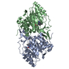
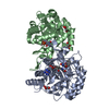
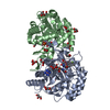
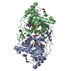
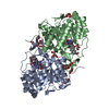
| Title | The R57A mutant of Lactococcus lactis dihydroorotate dehydrogenase A | ||||||
 Components Components | dihydroorotate dehydrogenase A | ||||||
 Keywords Keywords | OXIDOREDUCTASE / Homodimer / alpha-beta barrel / flavoprotein / orotate complex / mutant enzyme | ||||||
| Function / homology |  Function and homology information Function and homology informationdihydroorotate dehydrogenase (fumarate) / dihydroorotate dehydrogenase (fumarate) activity / 'de novo' UMP biosynthetic process / 'de novo' pyrimidine nucleobase biosynthetic process / cytoplasm Similarity search - Function | ||||||
| Biological species |  Lactococcus lactis (lactic acid bacteria) Lactococcus lactis (lactic acid bacteria) | ||||||
| Method |  X-RAY DIFFRACTION / SYNCHROTRON / MOLECULAR REPLACEMENT / Resolution: 1.7 Å X-RAY DIFFRACTION / SYNCHROTRON / MOLECULAR REPLACEMENT / Resolution: 1.7 Å | ||||||
 Authors Authors | Norager, S. / Arent, S. / Bjornberg, O. / Ottosen, M. / Lo Leggio, L. / Jensen, K.F. / Larsen, S. | ||||||
 Citation Citation | Journal: J.Biol.Chem. / Year: 2003 Title: Lactococcus lactis dihydroorotate dehydrogenase A mutants reveal important facets of the enzymatic function Authors: Norager, S. / Arent, S. / Bjornberg, O. / Ottosen, M. / Lo Leggio, L. / Jensen, K.F. / Larsen, S. #1: Journal: Biochemistry / Year: 1997Title: Active site of dihydroorotate dehydrogenase A from Lactococcus lactis investigated by chemical modification and mutagenesis Authors: Bjornberg, O. / Rowland, P. / Larsen, S. / Jensen, K.F. #2: Journal: Protein Sci. / Year: 1998Title: The crystal structure of Lactococcus lactis dihydroorotate dehydrogenase A complexed with the enzyme reaction product throws light on its enzymatic function Authors: Rowland, P. / Bjornberg, O. / Nielsen, F.S. / Jensen, K.F. / Larsen, S. #3: Journal: Structure / Year: 1997Title: The crystal structure of the flavin containing enzyme dihydroorotate dehydrogenase A from Lactococcus lactis Authors: Rowland, P. / Nielsen, F.S. / Jensen, K.F. / Larsen, S. #4: Journal: Protein Sci. / Year: 1996Title: Purification and characterisation of dihydroorotate dehydrogenase A from Lactococcus lactis, crystallisation and preliminary X-ray diffraction studies of the enzyme Authors: Nielsen, F.S. / Rowland, P. / Larsen, S. / Jensen, K.F. | ||||||
| History |
|
- Structure visualization
| Structure viewer | Molecule: MolmilJmol/JSmol |
|---|
- Downloads & links
-Download
| PDBx/mmCIF format | 1jqx.cif.gz | 154.6 KB | Display | PDBx/mmCIF format |
|---|---|---|---|---|
| PDB format | pdb1jqx.ent.gz | 118.5 KB | Display | PDB format |
| PDBx/mmJSON format | 1jqx.json.gz | Tree view | PDBx/mmJSON format | |
| Others |  Other downloads Other downloads |
-Validation report
| Arichive directory | https://data.pdbj.org/pub/pdb/validation_reports/jq/1jqxftp://data.pdbj.org/pub/pdb/validation_reports/jq/1jqx | HTTPS FTP |
|---|
-Related structure data
| Related structure data | 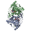 1jqvC 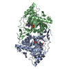 1jrbC  1jrcC  1jubC  1jueC  1ovdC 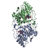 2dorS S: Starting model for refinement C: citing same article ( |
|---|---|
| Similar structure data |


-Links
 PDBj
PDBj- Assembly
| Deposited unit | 
| ||||||||
|---|---|---|---|---|---|---|---|---|---|
| 1 |
| ||||||||
| Unit cell |
| ||||||||
| Details | The assymetric unit contains the biological homodimer. |
-Components
-Protein , 1 types, 2 molecules AB
| #1: Protein | Mass: 34156.055 Da / Num. of mol.: 2 / Mutation: R57A Source method: isolated from a genetically manipulated source Source: (gene. exp.) Lactococcus lactis (lactic acid bacteria)Plasmid: pUHE23 / Production host: References: UniProt: P54321, UniProt: A2RJT9*PLUS, EC: 1.3.3.1 |
|---|
-Non-polymers , 5 types, 731 molecules 


| #2: Chemical |  Mass: 24.305 Da / Num. of mol.: 2 / Source method: obtained synthetically / Formula: Mg Mass: 24.305 Da / Num. of mol.: 2 / Source method: obtained synthetically / Formula: Mg#3: Chemical |  Mass: 456.344 Da / Num. of mol.: 2 / Source method: obtained synthetically / Formula: C17H21N4O9P Mass: 456.344 Da / Num. of mol.: 2 / Source method: obtained synthetically / Formula: C17H21N4O9P#4: Chemical |  Mass: 156.096 Da / Num. of mol.: 2 / Source method: obtained synthetically / Formula: C5H4N2O4 Mass: 156.096 Da / Num. of mol.: 2 / Source method: obtained synthetically / Formula: C5H4N2O4#5: Chemical |  Mass: 92.094 Da / Num. of mol.: 3 / Source method: obtained synthetically / Formula: C3H8O3 Mass: 92.094 Da / Num. of mol.: 3 / Source method: obtained synthetically / Formula: C3H8O3#6: Water | ChemComp-HOH / | Mass: 18.015 Da / Num. of mol.: 722 / Source method: isolated from a natural source / Formula: H2O |
|---|
-Experimental details
-Experiment
| Experiment | Method: X-RAY DIFFRACTION / Number of used crystals: 1 |
|---|
- Sample preparation
| Crystal | Density Matthews: 2.72 Å3/Da / Density % sol: 54.71 % |
|---|---|
| Crystal grow | Temperature: 293 K / Method: vapor diffusion, hanging drop Details: PEG 6K, Na-acetate, TRIS-HCl, VAPOR DIFFUSION, HANGING DROP, temperature 293K |
-Data collection
| Diffraction | Mean temperature: 120 K |
|---|---|
| Diffraction source | Source: SYNCHROTRON / Site: ESRF  / Beamline: ID14-2 / Wavelength: 0.933 Å / Beamline: ID14-2 / Wavelength: 0.933 Å |
| Detector | Type: ADSC QUANTUM 4 / Detector: CCD / Date: Mar 26, 2001 |
| Radiation | Monochromator: DIAMOND / Protocol: SINGLE WAVELENGTH / Monochromatic (M) / Laue (L): M / Scattering type: x-ray |
| Radiation wavelength | Wavelength: 0.933 Å / Relative weight: 1 |
| Reflection | Resolution: 1.7→25 Å / Num. all: 679947 / Num. obs: 80319 / % possible obs: 99.8 % / Observed criterion σ(F): 0 / Observed criterion σ(I): 0 / Redundancy: 4 % / Biso Wilson estimate: 17.4 Å2 / Rmerge(I) obs: 0.047 / Net I/σ(I): 24 |
| Reflection shell | Resolution: 1.7→1.73 Å / Rmerge(I) obs: 0.151 / Mean I/σ(I) obs: 6.8 / Num. unique all: 3949 / % possible all: 97.4 |
- Processing
| Software |
| |||||||||||||||||||||||||
|---|---|---|---|---|---|---|---|---|---|---|---|---|---|---|---|---|---|---|---|---|---|---|---|---|---|---|
| Refinement | Method to determine structure: MOLECULAR REPLACEMENT Starting model: Lactococcus lactis DHODA PDB ID 2DOR Resolution: 1.7→25 Å / Rfactor Rfree error: 0.002 / Isotropic thermal model: isotropic / Cross valid method: THROUGHOUT / σ(F): 0 / σ(I): 0 / Stereochemistry target values: Engh & Huber
| |||||||||||||||||||||||||
| Solvent computation | Solvent model: FLAT MODEL / Bsol: 54.8305 Å2 / ksol: 0.368323 e/Å3 | |||||||||||||||||||||||||
| Displacement parameters | Biso mean: 19 Å2 | |||||||||||||||||||||||||
| Refine analyze |
| |||||||||||||||||||||||||
| Refinement step | Cycle: LAST / Resolution: 1.7→25 Å
| |||||||||||||||||||||||||
| Refine LS restraints |
| |||||||||||||||||||||||||
| LS refinement shell | Resolution: 1.7→1.81 Å / Rfactor Rfree error: 0.006
|
