 Movie
Movie Controller

[English] 日本語
 Yorodumi
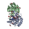
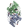
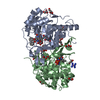
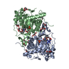
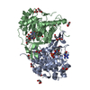
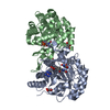
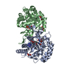
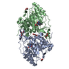
- PDB-2dor: DIHYDROOROTATE DEHYDROGENASE A FROM LACTOCOCCUS LACTIS COMPLEXED ... -
+ Open data

- Basic information
| Entry | Database: PDB / ID: 2dor | ||||||
|---|---|---|---|---|---|---|---|
| Title | DIHYDROOROTATE DEHYDROGENASE A FROM LACTOCOCCUS LACTIS COMPLEXED WITH OROTATE | ||||||
 Components Components | DIHYDROOROTATE DEHYDROGENASE A | ||||||
 Keywords Keywords | OXIDOREDUCTASE / PYRIMIDINE NUCLEOTIDE BIOSYNTHESIS | ||||||
| Function / homology |  Function and homology information Function and homology informationdihydroorotate dehydrogenase (fumarate) / dihydroorotate dehydrogenase (fumarate) activity / 'de novo' UMP biosynthetic process / 'de novo' pyrimidine nucleobase biosynthetic process / cytoplasm Similarity search - Function | ||||||
| Biological species |  Lactococcus lactis (lactic acid bacteria) Lactococcus lactis (lactic acid bacteria) | ||||||
| Method |  X-RAY DIFFRACTION / DIFFERENCE MAP / Resolution: 2 Å X-RAY DIFFRACTION / DIFFERENCE MAP / Resolution: 2 Å | ||||||
 Authors Authors | Rowland, P. / Larsen, S. | ||||||
 Citation Citation | Journal: Protein Sci. / Year: 1998 Title: The crystal structure of Lactococcus lactis dihydroorotate dehydrogenase A complexed with the enzyme reaction product throws light on its enzymatic function. Authors: Rowland, P. / Bjornberg, O. / Nielsen, F.S. / Jensen, K.F. / Larsen, S. | ||||||
| History |
|
- Structure visualization
| Structure viewer | Molecule: MolmilJmol/JSmol |
|---|
- Downloads & links
-Download
| PDBx/mmCIF format | 2dor.cif.gz | 138.9 KB | Display | PDBx/mmCIF format |
|---|---|---|---|---|
| PDB format | pdb2dor.ent.gz | 108.3 KB | Display | PDB format |
| PDBx/mmJSON format | 2dor.json.gz | Tree view | PDBx/mmJSON format | |
| Others |  Other downloads Other downloads |
-Validation report
| Arichive directory | https://data.pdbj.org/pub/pdb/validation_reports/do/2dorftp://data.pdbj.org/pub/pdb/validation_reports/do/2dor | HTTPS FTP |
|---|
-Related structure data
| Similar structure data |
|---|


-Links
 PDBj
PDBj- Assembly
| Deposited unit | 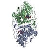
| ||||||||
|---|---|---|---|---|---|---|---|---|---|
| 1 |
| ||||||||
| Unit cell |
| ||||||||
| Noncrystallographic symmetry (NCS) | NCS oper: (Code: given Matrix: (0.8728, 0.0049, 0.4881), Vector: |
-Components
| #1: Protein | Mass: 34242.168 Da / Num. of mol.: 2 Source method: isolated from a genetically manipulated source Source: (gene. exp.) Lactococcus lactis (lactic acid bacteria)Production host: References: UniProt: P54321, UniProt: A2RJT9*PLUS, EC: 1.3.3.1 #2: Chemical |   Mass: 456.344 Da / Num. of mol.: 2 / Source method: obtained synthetically / Formula: C17H21N4O9P Mass: 456.344 Da / Num. of mol.: 2 / Source method: obtained synthetically / Formula: C17H21N4O9P#3: Chemical | 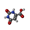  Mass: 156.096 Da / Num. of mol.: 2 / Source method: obtained synthetically / Formula: C5H4N2O4 Mass: 156.096 Da / Num. of mol.: 2 / Source method: obtained synthetically / Formula: C5H4N2O4#4: Water | ChemComp-HOH / |  Mass: 18.015 Da / Num. of mol.: 346 / Source method: isolated from a natural source / Formula: H2O Mass: 18.015 Da / Num. of mol.: 346 / Source method: isolated from a natural source / Formula: H2O |
|---|
-Experimental details
-Experiment
| Experiment | Method: X-RAY DIFFRACTION / Number of used crystals: 1 |
|---|
- Sample preparation
| Crystal | Density Matthews: 2.81 Å3/Da / Density % sol: 56 % | ||||||||||||||||||||||||||||||
|---|---|---|---|---|---|---|---|---|---|---|---|---|---|---|---|---|---|---|---|---|---|---|---|---|---|---|---|---|---|---|---|
| Crystal grow | pH: 8.5 Details: PROTEIN WAS CRYSTALLISED FROM A SOLUTION CONTAINING 30% PEG 6000, 0.2M SODIUM ACETATE AND 0.1M TRIS-HCL, PH 8.5. | ||||||||||||||||||||||||||||||
| Crystal | *PLUS | ||||||||||||||||||||||||||||||
| Crystal grow | *PLUS Method: vapor diffusion, hanging drop | ||||||||||||||||||||||||||||||
| Components of the solutions | *PLUS
|
-Data collection
| Diffraction | Mean temperature: 288 K |
|---|---|
| Diffraction source | Source: ROTATING ANODE / Type: RIGAKU RUH2R / Wavelength: 1.5418 |
| Detector | Type: RIGAKU RAXIS II / Detector: IMAGE PLATE / Date: Nov 1, 1995 / Details: COLLIMATOR |
| Radiation | Monochromator: GRAPHITE(002) / Monochromatic (M) / Laue (L): M / Scattering type: x-ray |
| Radiation wavelength | Wavelength: 1.5418 Å / Relative weight: 1 |
| Reflection | Resolution: 2→25 Å / Num. obs: 50309 / % possible obs: 99.3 % / Observed criterion σ(I): -3 / Redundancy: 3.5 % / Biso Wilson estimate: 24.2 Å2 / Rsym value: 0.061 / Net I/σ(I): 18.7 |
| Reflection shell | Resolution: 2→2.03 Å / Redundancy: 2.6 % / Mean I/σ(I) obs: 3.6 / Rsym value: 0.25 / % possible all: 90.5 |
| Reflection | *PLUS Num. measured all: 179226 / Rmerge(I) obs: 0.061 |
| Reflection shell | *PLUS % possible obs: 90.5 % / Rmerge(I) obs: 0.25 |
- Processing
| Software |
| ||||||||||||||||||||||||||||||||||||||||||||||||||||||||||||
|---|---|---|---|---|---|---|---|---|---|---|---|---|---|---|---|---|---|---|---|---|---|---|---|---|---|---|---|---|---|---|---|---|---|---|---|---|---|---|---|---|---|---|---|---|---|---|---|---|---|---|---|---|---|---|---|---|---|---|---|---|---|
| Refinement | Method to determine structure: DIFFERENCE MAP / Resolution: 2→25 Å / Rfactor Rfree error: 0.004 / Isotropic thermal model: RESTRAINED / Cross valid method: FREE R
| ||||||||||||||||||||||||||||||||||||||||||||||||||||||||||||
| Displacement parameters | Biso mean: 27.3 Å2 | ||||||||||||||||||||||||||||||||||||||||||||||||||||||||||||
| Refine analyze | Luzzati d res low obs: 25 Å / Luzzati sigma a obs: 0.2 Å | ||||||||||||||||||||||||||||||||||||||||||||||||||||||||||||
| Refinement step | Cycle: LAST / Resolution: 2→25 Å
| ||||||||||||||||||||||||||||||||||||||||||||||||||||||||||||
| Refine LS restraints |
| ||||||||||||||||||||||||||||||||||||||||||||||||||||||||||||
| LS refinement shell | Resolution: 2→2.03 Å / Rfactor Rfree error: 0.03 / Total num. of bins used: 20
| ||||||||||||||||||||||||||||||||||||||||||||||||||||||||||||
| Xplor file |
| ||||||||||||||||||||||||||||||||||||||||||||||||||||||||||||
| Software | *PLUS Name: X-PLOR / Version: 3.1 / Classification: refinement | ||||||||||||||||||||||||||||||||||||||||||||||||||||||||||||
| Refinement | *PLUS Num. reflection all: 50283 / Num. reflection obs: 48238 | ||||||||||||||||||||||||||||||||||||||||||||||||||||||||||||
| Solvent computation | *PLUS | ||||||||||||||||||||||||||||||||||||||||||||||||||||||||||||
| Displacement parameters | *PLUS | ||||||||||||||||||||||||||||||||||||||||||||||||||||||||||||
| Refine LS restraints | *PLUS
|
