- PDB-2bsh: Crystal structure of the type III secretion chaperone SycT from Y... -
+
Open data
ID or keywords:
Loading...
-
Basic information
Entry
Database: PDB / ID: 2bsh
Title
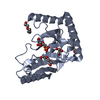
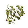
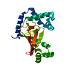
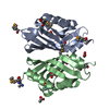
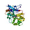
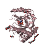
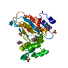
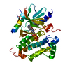
Crystal structure of the type III secretion chaperone SycT from Yersinia enterocolitica (crystal form 2)
Components
SYCT
Keywords
CHAPERONE / TYPE III SECRETION / YERSINIA / EFFECTOR / YOPT
Function / homology
Tir chaperone protein (CesT) family / Tir chaperone protein (CesT) family / Yope Regulator; Chain: A, - #10 / Yope Regulator; Chain: A, / protein secretion by the type III secretion system / 2-Layer Sandwich / Alpha Beta / Chaperone protein SycT / Chaperone protein SycT
Function and homology information
Biological species
YERSINIA ENTEROCOLITICA (bacteria)
Method
X-RAY DIFFRACTION / SYNCHROTRON / SAD / Resolution: 1.9 Å
 Movie
Movie Controller

 Yorodumi
 Open data
 Basic information
 Components
 Keywords
 Function and homology information
 YERSINIA ENTEROCOLITICA (bacteria)
 X-RAY DIFFRACTION /
  Authors
 Citation
 Structure visualization
 Downloads & links
 Other downloads
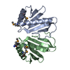


 PDBj
PDBj
 Assembly
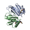
 Mass: 18.015 Da / Num. of mol.: 139 / Source method: isolated from a natural source / Formula: H2O
 Sample preparation
 / Beamline: 14.1 / Wavelength: 0.97957
  Processing