 Movie
Movie Controller

[English] 日本語
 Yorodumi
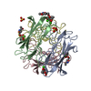
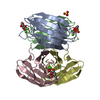
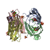
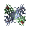
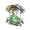
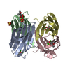
- PDB-2boi: 1.1A Structure of Chromobacterium Violaceum Lectin CV2L in Comple... -
+ Open data

- Basic information
| Entry | Database: PDB / ID: 2boi | ||||||
|---|---|---|---|---|---|---|---|
| Title | 1.1A Structure of Chromobacterium Violaceum Lectin CV2L in Complex with alpha-methyl-fucoside | ||||||
 Components Components | CV-IIL LECTIN | ||||||
 Keywords Keywords | LECTIN / FUCOSE / CHROMOBACTERIUM VIOLACEUM / PSEUDOMONAS AERUGINOSA | ||||||
| Function / homology |  Function and homology information Function and homology information | ||||||
| Biological species |  CHROMOBACTERIUM VIOLACEUM (bacteria) CHROMOBACTERIUM VIOLACEUM (bacteria) | ||||||
| Method |  X-RAY DIFFRACTION / SYNCHROTRON / MOLECULAR REPLACEMENT / Resolution: 1.1 Å X-RAY DIFFRACTION / SYNCHROTRON / MOLECULAR REPLACEMENT / Resolution: 1.1 Å | ||||||
 Authors Authors | Pokorna, M. / Cioci, G. / Perret, S. / Rebuffet, E. / Adam, J. / Gilboa-Garber, N. / Mitchell, E.P. / Imberty, A. / Wimmerova, M. | ||||||
 Citation Citation | Journal: Biochemistry / Year: 2006 Title: Unusual Entropy Driven Affinity of Chromobacter Violaceum Lectin Cv-Iil Towards Fucose and Mannose Authors: Pokorna, M. / Cioci, G. / Perret, S. / Rebuffet, E. / Kostlanova, N. / Adam, J. / Gilboa-Garber, N. / Mitchell, E.P. / Imberty, A. / Wimmerova, M. | ||||||
| History |
| ||||||
| Remark 700 | SHEET DETERMIMATION METHOD: PROVIDED BY DEPOSITOR |
- Structure visualization
| Structure viewer | Molecule: MolmilJmol/JSmol |
|---|
- Downloads & links
-Download
| PDBx/mmCIF format | 2boi.cif.gz | 117.6 KB | Display | PDBx/mmCIF format |
|---|---|---|---|---|
| PDB format | pdb2boi.ent.gz | 90.5 KB | Display | PDB format |
| PDBx/mmJSON format | 2boi.json.gz | Tree view | PDBx/mmJSON format | |
| Others |  Other downloads Other downloads |
-Validation report
| Arichive directory | https://data.pdbj.org/pub/pdb/validation_reports/bo/2boiftp://data.pdbj.org/pub/pdb/validation_reports/bo/2boi | HTTPS FTP |
|---|
-Related structure data
| Related structure data |  2bv4C  1gztS C: citing same article ( S: Starting model for refinement |
|---|---|
| Similar structure data |
-Links
 PDBj
PDBj- Assembly
| Deposited unit | 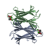
| ||||||||
|---|---|---|---|---|---|---|---|---|---|
| 1 | 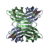
| ||||||||
| Unit cell |
|
-Components
| #1: Protein | Mass: 11848.111 Da / Num. of mol.: 2 Source method: isolated from a genetically manipulated source Source: (gene. exp.) CHROMOBACTERIUM VIOLACEUM (bacteria) / Plasmid: PET25(B) / Production host: #2: Chemical | ChemComp-CA /   Mass: 40.078 Da / Num. of mol.: 4 / Source method: obtained synthetically / Formula: Ca Mass: 40.078 Da / Num. of mol.: 4 / Source method: obtained synthetically / Formula: Ca#3: Sugar | 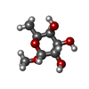  Type: L-saccharide / Mass: 178.183 Da / Num. of mol.: 2 Type: L-saccharide / Mass: 178.183 Da / Num. of mol.: 2Source method: isolated from a genetically manipulated source Formula: C7H14O5 #4: Water | ChemComp-HOH / |  Mass: 18.015 Da / Num. of mol.: 333 / Source method: isolated from a natural source / Formula: H2O Mass: 18.015 Da / Num. of mol.: 333 / Source method: isolated from a natural source / Formula: H2O |
|---|
-Experimental details
-Experiment
| Experiment | Method: X-RAY DIFFRACTION / Number of used crystals: 1 |
|---|
- Sample preparation
| Crystal | Density Matthews: 2.2 Å3/Da / Density % sol: 45 % Description: MOLREP WAS USED WITH MODEL 1GZT TO FIND THE POSITIONS OF 4 CA ATOMS IN THE A.U. PHASING WAS PERFORMED BY ACORN STARTING FROM THE POSITIONS OF THESE CA ATOMS. |
|---|---|
| Crystal grow | Details: PEG 8000 10%, 0.1 M (NH4)2SO4 PH 5 |
-Data collection
| Diffraction | Mean temperature: 100 K |
|---|---|
| Diffraction source | Source: SYNCHROTRON / Site: ESRF  / Beamline: ID14-2 / Wavelength: 0.933 / Beamline: ID14-2 / Wavelength: 0.933 |
| Detector | Type: ADSC CCD / Detector: CCD / Date: Jul 15, 2004 / Details: MIRRORS |
| Radiation | Monochromator: DIAMOND AND GE DOUBLE CRYSTAL / Protocol: SINGLE WAVELENGTH / Monochromatic (M) / Laue (L): M / Scattering type: x-ray |
| Radiation wavelength | Wavelength: 0.933 Å / Relative weight: 1 |
| Reflection | Resolution: 1.1→34.38 Å / Num. obs: 87549 / % possible obs: 100 % / Observed criterion σ(I): 0 / Redundancy: 8.14 % / Rmerge(I) obs: 0.07 / Net I/σ(I): 5.36 |
| Reflection shell | Resolution: 1.1→1.13 Å / Redundancy: 7.04 % / Rmerge(I) obs: 0.3 / Mean I/σ(I) obs: 2.34 / % possible all: 100 |
- Processing
| Software |
| ||||||||||||||||||||||||||||||||||||||||||||||||||||||||||||||||||||||||||||||||||||||||||||||||||||||||||||||||||||||||||||||||||||||||||||||||||||||||||||||||||||||||||||||||||||||
|---|---|---|---|---|---|---|---|---|---|---|---|---|---|---|---|---|---|---|---|---|---|---|---|---|---|---|---|---|---|---|---|---|---|---|---|---|---|---|---|---|---|---|---|---|---|---|---|---|---|---|---|---|---|---|---|---|---|---|---|---|---|---|---|---|---|---|---|---|---|---|---|---|---|---|---|---|---|---|---|---|---|---|---|---|---|---|---|---|---|---|---|---|---|---|---|---|---|---|---|---|---|---|---|---|---|---|---|---|---|---|---|---|---|---|---|---|---|---|---|---|---|---|---|---|---|---|---|---|---|---|---|---|---|---|---|---|---|---|---|---|---|---|---|---|---|---|---|---|---|---|---|---|---|---|---|---|---|---|---|---|---|---|---|---|---|---|---|---|---|---|---|---|---|---|---|---|---|---|---|---|---|---|---|
| Refinement | Method to determine structure: MOLECULAR REPLACEMENT Starting model: PDB ENTRY 1GZT Resolution: 1.1→46.47 Å / Cor.coef. Fo:Fc: 0.983 / Cor.coef. Fo:Fc free: 0.979 / SU B: 0.541 / SU ML: 0.012 / Cross valid method: THROUGHOUT / ESU R: 0.021 / ESU R Free: 0.021 / Stereochemistry target values: MAXIMUM LIKELIHOOD / Details: HYDROGENS HAVE BEEN ADDED IN THE RIDING POSITIONS.
| ||||||||||||||||||||||||||||||||||||||||||||||||||||||||||||||||||||||||||||||||||||||||||||||||||||||||||||||||||||||||||||||||||||||||||||||||||||||||||||||||||||||||||||||||||||||
| Solvent computation | Ion probe radii: 0.8 Å / Shrinkage radii: 0.8 Å / VDW probe radii: 1.2 Å / Solvent model: MASK | ||||||||||||||||||||||||||||||||||||||||||||||||||||||||||||||||||||||||||||||||||||||||||||||||||||||||||||||||||||||||||||||||||||||||||||||||||||||||||||||||||||||||||||||||||||||
| Refinement step | Cycle: LAST / Resolution: 1.1→46.47 Å
| ||||||||||||||||||||||||||||||||||||||||||||||||||||||||||||||||||||||||||||||||||||||||||||||||||||||||||||||||||||||||||||||||||||||||||||||||||||||||||||||||||||||||||||||||||||||
| Refine LS restraints |
|