 Movie
Movie Controller

[English] 日本語
 Yorodumi
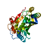
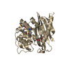
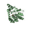
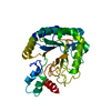
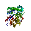
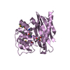
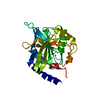
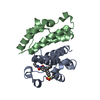
- PDB-2bof: Catalytic domain of endo-1,4-glucanase Cel6A mutant Y73S from The... -
+ Open data

- Basic information
| Entry | Database: PDB / ID: 2bof | |||||||||
|---|---|---|---|---|---|---|---|---|---|---|
| Title | Catalytic domain of endo-1,4-glucanase Cel6A mutant Y73S from Thermobifida fusca in complex with cellotetrose | |||||||||
 Components Components | ENDOGLUCANASE E-2 | |||||||||
 Keywords Keywords | HYDROLASE / ENDOGLUCANASE / THERMOBIFIDA FUSCA / TIM A/B FOLD / GLYCOSIDE HYDROLASE FAMILY 6 / METHYL CELLOBIOSYL-4-THIO-BETA- CELLOBIOSIDE | |||||||||
| Function / homology |  Function and homology information Function and homology informationcellulase / cellulase activity / polysaccharide binding / cellulose catabolic process Similarity search - Function | |||||||||
| Biological species |   THERMOMONOSPORA FUSCA (bacteria) THERMOMONOSPORA FUSCA (bacteria) | |||||||||
| Method |  X-RAY DIFFRACTION / SYNCHROTRON / MOLECULAR REPLACEMENT / Resolution: 1.64 Å X-RAY DIFFRACTION / SYNCHROTRON / MOLECULAR REPLACEMENT / Resolution: 1.64 Å | |||||||||
 Authors Authors | Larsson, A.M. / Bergfors, T. / Dultz, E. / Irwin, D.C. / Roos, A. / Driguez, H. / Wilson, D.B. / Jones, T.A. | |||||||||
 Citation Citation | Journal: Biochemistry / Year: 2005 Title: Crystal Structure of Thermobifida Fusca Endoglucanase Cel6A in Complex with Substrate and Inhibitor: The Role of Tyrosine Y73 in Substrate Ring Distortion. Authors: Larsson, A.M. / Bergfors, T. / Dultz, E. / Irwin, D.C. / Roos, A. / Driguez, H. / Wilson, D.B. / Jones, T.A. | |||||||||
| History |
|
- Structure visualization
| Structure viewer | Molecule: MolmilJmol/JSmol |
|---|
- Downloads & links
-Download
| PDBx/mmCIF format | 2bof.cif.gz | 73.2 KB | Display | PDBx/mmCIF format |
|---|---|---|---|---|
| PDB format | pdb2bof.ent.gz | 53.2 KB | Display | PDB format |
| PDBx/mmJSON format | 2bof.json.gz | Tree view | PDBx/mmJSON format | |
| Others |  Other downloads Other downloads |
-Validation report
| Arichive directory | https://data.pdbj.org/pub/pdb/validation_reports/bo/2bofftp://data.pdbj.org/pub/pdb/validation_reports/bo/2bof | HTTPS FTP |
|---|
-Related structure data
| Related structure data | 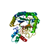 2bodC 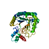 2boeC 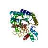 2bogC 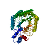 1tmlS S: Starting model for refinement C: citing same article ( |
|---|---|
| Similar structure data |


-Links
 PDBj
PDBj


- Assembly
| Deposited unit | 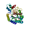
| ||||||||
|---|---|---|---|---|---|---|---|---|---|
| 1 |
| ||||||||
| Unit cell |
|
-Components
| #1: Protein | Mass: 30360.695 Da / Num. of mol.: 1 / Fragment: CATALYTIC DOMAIN, RESIDUES 32-317 / Mutation: YES Source method: isolated from a genetically manipulated source Source: (gene. exp.) THERMOMONOSPORA FUSCA (bacteria) / Strain: YX / Plasmid: POS12 / Production host: | ||
|---|---|---|---|
| #2: Polysaccharide | beta-D-glucopyranose-(1-4)-beta-D-glucopyranose-(1-4)-beta-D-glucopyranose-(1-4)-beta-D-glucopyranose / beta-cellotetraose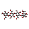  Source method: isolated from a genetically manipulated source Details: oligosaccharide / References: beta-cellotetraose | ||
| #3: Water | ChemComp-HOH /  Mass: 18.015 Da / Num. of mol.: 273 / Source method: isolated from a natural source / Formula: H2O Mass: 18.015 Da / Num. of mol.: 273 / Source method: isolated from a natural source / Formula: H2O | ||
| Compound details | ENGINEERED| Has protein modification | Y | |
-Experimental details
-Experiment
| Experiment | Method: X-RAY DIFFRACTION / Number of used crystals: 1 |
|---|
- Sample preparation
| Crystal | Density Matthews: 2 Å3/Da / Density % sol: 36.6 % |
|---|---|
| Crystal grow | Details: 20-30% POLYETHYLENGLYCOL 4000 AND 0.15-0.34 M SODIUM MALONATE, PH 4.0 |
-Data collection
| Diffraction | Mean temperature: 100 K |
|---|---|
| Diffraction source | Source: SYNCHROTRON / Site: MAX II  / Beamline: I711 / Wavelength: 1.089 / Beamline: I711 / Wavelength: 1.089 |
| Detector | Type: MARRESEARCH / Detector: CCD / Date: May 21, 2003 / Details: SI MIRROR COATED WITH PLATINUM |
| Radiation | Monochromator: ASYMMETRICALLY CUT SINGLE SI(111) / Protocol: SINGLE WAVELENGTH / Monochromatic (M) / Laue (L): M / Scattering type: x-ray |
| Radiation wavelength | Wavelength: 1.089 Å / Relative weight: 1 |
| Reflection | Resolution: 1.64→52 Å / Num. obs: 28960 / % possible obs: 99.6 % / Observed criterion σ(I): 6 / Redundancy: 4.1 % / Rmerge(I) obs: 0.07 / Rsym value: 0.061 / Net I/σ(I): 15.6 |
| Reflection shell | Resolution: 1.64→1.73 Å / Redundancy: 3.9 % / Rmerge(I) obs: 0.2 / Mean I/σ(I) obs: 6.3 / Rsym value: 0.174 / % possible all: 97.1 |
- Processing
| Software |
| ||||||||||||||||||||||||||||||||||||||||||||||||||||||||||||||||||||||||||||||||||||||||||||||||||||||||||||||||||||||||||||||||||||||||||||||||||||||||||||||||||||||||||||||||||||||
|---|---|---|---|---|---|---|---|---|---|---|---|---|---|---|---|---|---|---|---|---|---|---|---|---|---|---|---|---|---|---|---|---|---|---|---|---|---|---|---|---|---|---|---|---|---|---|---|---|---|---|---|---|---|---|---|---|---|---|---|---|---|---|---|---|---|---|---|---|---|---|---|---|---|---|---|---|---|---|---|---|---|---|---|---|---|---|---|---|---|---|---|---|---|---|---|---|---|---|---|---|---|---|---|---|---|---|---|---|---|---|---|---|---|---|---|---|---|---|---|---|---|---|---|---|---|---|---|---|---|---|---|---|---|---|---|---|---|---|---|---|---|---|---|---|---|---|---|---|---|---|---|---|---|---|---|---|---|---|---|---|---|---|---|---|---|---|---|---|---|---|---|---|---|---|---|---|---|---|---|---|---|---|---|
| Refinement | Method to determine structure: MOLECULAR REPLACEMENT Starting model: PDB ENTRY 1TML Resolution: 1.64→51.99 Å / Cor.coef. Fo:Fc: 0.966 / Cor.coef. Fo:Fc free: 0.951 / SU B: 1.592 / SU ML: 0.056 / Cross valid method: THROUGHOUT / ESU R: 0.096 / ESU R Free: 0.092 / Stereochemistry target values: MAXIMUM LIKELIHOOD / Details: HYDROGENS HAVE BEEN ADDED IN THE RIDING POSITIONS.
| ||||||||||||||||||||||||||||||||||||||||||||||||||||||||||||||||||||||||||||||||||||||||||||||||||||||||||||||||||||||||||||||||||||||||||||||||||||||||||||||||||||||||||||||||||||||
| Solvent computation | Ion probe radii: 0.8 Å / Shrinkage radii: 0.8 Å / VDW probe radii: 1.4 Å / Solvent model: BABINET MODEL WITH MASK | ||||||||||||||||||||||||||||||||||||||||||||||||||||||||||||||||||||||||||||||||||||||||||||||||||||||||||||||||||||||||||||||||||||||||||||||||||||||||||||||||||||||||||||||||||||||
| Displacement parameters | Biso mean: 12.62 Å2
| ||||||||||||||||||||||||||||||||||||||||||||||||||||||||||||||||||||||||||||||||||||||||||||||||||||||||||||||||||||||||||||||||||||||||||||||||||||||||||||||||||||||||||||||||||||||
| Refinement step | Cycle: LAST / Resolution: 1.64→51.99 Å
| ||||||||||||||||||||||||||||||||||||||||||||||||||||||||||||||||||||||||||||||||||||||||||||||||||||||||||||||||||||||||||||||||||||||||||||||||||||||||||||||||||||||||||||||||||||||
| Refine LS restraints |
|