 ムービー
ムービー コントローラー


+ データを開く

- 基本情報
基本情報
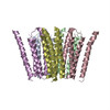
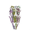
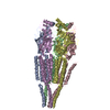
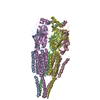
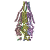
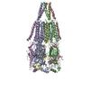
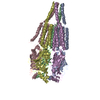
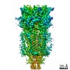
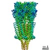
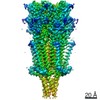
| 登録情報 | データベース: PDB / ID: 2bg9 | ||||||
|---|---|---|---|---|---|---|---|
| タイトル | REFINED STRUCTURE OF THE NICOTINIC ACETYLCHOLINE RECEPTOR AT 4A RESOLUTION. | ||||||
 要素 要素 |
| ||||||
 キーワード キーワード | ION CHANNEL/RECEPTOR / ACETYLCHOLINE RECEPTOR / ION CHANNEL / ION TRANSPORT / POSTSYNAPTIC MEMBRANE / ION CHANNEL-RECEPTOR complex | ||||||
| 機能・相同性 |  機能・相同性情報 機能・相同性情報acetylcholine-gated channel complex / acetylcholine-gated monoatomic cation-selective channel activity / acetylcholine receptor signaling pathway / transmembrane signaling receptor activity / postsynaptic membrane 類似検索 - 分子機能 | ||||||
| 生物種 |  | ||||||
| 手法 | 電子顕微鏡法 / らせん対称体再構成法 / クライオ電子顕微鏡法 / 解像度: 4 Å | ||||||
 データ登録者 データ登録者 | Unwin, N. | ||||||
 引用 引用 | ジャーナル: J Mol Biol / 年: 2005 タイトル: Refined structure of the nicotinic acetylcholine receptor at 4A resolution. 著者: Nigel Unwin /  要旨: We present a refined model of the membrane-associated Torpedo acetylcholine (ACh) receptor at 4A resolution. An improved experimental density map was obtained from 342 electron images of helical ...We present a refined model of the membrane-associated Torpedo acetylcholine (ACh) receptor at 4A resolution. An improved experimental density map was obtained from 342 electron images of helical tubes, and the refined structure was derived to an R-factor of 36.7% (R(free) 37.9%) by standard crystallographic methods, after placing the densities corresponding to a single molecule into an artificial unit cell. The agreement between experimental and calculated phases along the helical layer-lines was used to monitor progress in the refinement and to give an independent measure of the accuracy. The atomic model allowed a detailed description of the whole receptor in the closed-channel form, including the ligand-binding and intracellular domains, which have not previously been interpreted at a chemical level. We confirm that the two ligand-binding alpha subunits have a different extended conformation from the three other subunits in the closed channel, and identify several interactions on both pairs of subunit interfaces, and within the alpha subunits, which may be responsible for their "distorted" structures. The ACh-coordinating amino acid side-chains of the alpha subunits are far apart in the closed channel, indicating that a localised rearrangement, involving closure of loops B and C around the bound ACh molecule, occurs upon activation. A comparison of the structure of the alpha subunit with that of AChBP having ligand present, suggests how the localised rearrangement overcomes the distortions and initiates the rotational movements associated with opening of the channel. Both vestibules of the channel are strongly electronegative, providing a cation-stabilising environment at either entrance of the membrane pore. Access to the pore on the intracellular side is further influenced by narrow lateral windows, which would be expected to screen out electrostatically ions of the wrong charge and size. #1: ジャーナル: Nature / 年: 2003タイトル: Structure and gating mechanism of the acetylcholine receptor pore. 著者: Atsuo Miyazawa / Yoshinori Fujiyoshi / Nigel Unwin /  要旨: The nicotinic acetylcholine receptor controls electrical signalling between nerve and muscle cells by opening and closing a gated, membrane-spanning pore. Here we present an atomic model of the ...The nicotinic acetylcholine receptor controls electrical signalling between nerve and muscle cells by opening and closing a gated, membrane-spanning pore. Here we present an atomic model of the closed pore, obtained by electron microscopy of crystalline postsynaptic membranes. The pore is shaped by an inner ring of 5 alpha-helices, which curve radially to create a tapering path for the ions, and an outer ring of 15 alpha-helices, which coil around each other and shield the inner ring from the lipids. The gate is a constricting hydrophobic girdle at the middle of the lipid bilayer, formed by weak interactions between neighbouring inner helices. When acetylcholine enters the ligand-binding domain, it triggers rotations of the protein chains on opposite sides of the entrance to the pore. These rotations are communicated through the inner helices, and open the pore by breaking the girdle apart. | ||||||
| 履歴 |
| ||||||
| Remark 650 | HELIX DETERMINATION METHOD: AUTHOR PROVIDED. | ||||||
| Remark 700 | SHEET THE SHEET STRUCTURE OF THIS MOLECULE IS BIFURCATED. IN ORDER TO REPRESENT THIS FEATURE IN ... SHEET THE SHEET STRUCTURE OF THIS MOLECULE IS BIFURCATED. IN ORDER TO REPRESENT THIS FEATURE IN THE SHEET RECORDS BELOW, TWO SHEETS ARE DEFINED. |
- 構造の表示
構造の表示
| ムービー |
ムービービューア |
|---|---|
| 構造ビューア | 分子: MolmilJmol/JSmol |
- ダウンロードとリンク
-ダウンロード
| PDBx/mmCIF形式 | 2bg9.cif.gz | 331.2 KB | 表示 | PDBx/mmCIF形式 |
|---|---|---|---|---|
| PDB形式 | pdb2bg9.ent.gz | 257.5 KB | 表示 | PDB形式 |
| PDBx/mmJSON形式 | 2bg9.json.gz | ツリー表示 | PDBx/mmJSON形式 | |
| その他 |  その他のダウンロード その他のダウンロード |
-検証レポート
| 文書・要旨 | 2bg9_validation.pdf.gz | 410.6 KB | 表示 | wwPDB検証レポート |
|---|---|---|---|---|
| 文書・詳細版 | 2bg9_full_validation.pdf.gz | 860.8 KB | 表示 | |
| XML形式データ | 2bg9_validation.xml.gz | 104.2 KB | 表示 | |
| CIF形式データ | 2bg9_validation.cif.gz | 143.6 KB | 表示 | |
| アーカイブディレクトリ | https://data.pdbj.org/pub/pdb/validation_reports/bg/2bg9ftp://data.pdbj.org/pub/pdb/validation_reports/bg/2bg9 | HTTPS FTP |
-関連構造データ
| 関連構造データ | |
|---|---|
| 類似構造データ |


-リンク
 PDBj
PDBj

- 集合体
集合体
| 登録構造単位 | 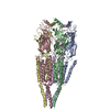
|
|---|---|
| 1 |
|
-要素
| #1: タンパク質 | 分子量: 42501.316 Da / 分子数: 2 / 由来タイプ: 天然 / 由来: (天然) #2: タンパク質 | | 分子量: 42167.359 Da / 分子数: 1 / 由来タイプ: 天然 / 由来: (天然) #3: タンパク質 | | 分子量: 42280.695 Da / 分子数: 1 / 由来タイプ: 天然 / 由来: (天然) #4: タンパク質 | | 分子量: 42299.496 Da / 分子数: 1 / 由来タイプ: 天然 / 詳細: INHABITS THE EUROPEAN COAST / 由来: (天然) Has protein modification | Y | |
|---|
-実験情報
-実験
| 実験 | 手法: 電子顕微鏡法 |
|---|---|
| EM実験 | 試料の集合状態: HELICAL ARRAY / 3次元再構成法: らせん対称体再構成法 |
- 試料調製
試料調製
| 構成要素 | 名称: NICOTINIC ACETYLCHOLINE RECEPTOR in postsynaptic membrane タイプ: COMPLEX |
|---|---|
| 緩衝液 | 名称: 100MM SODIUM CACODYLATE / pH: 6.8 / 詳細: 100MM SODIUM CACODYLATE |
| 試料 | 包埋: NO / シャドウイング: NO / 染色: NO / 凍結: YES |
| 試料支持 | 詳細: HOLEY CARBON |
| 急速凍結 | 詳細: LIQUID ETHANE |
- 電子顕微鏡撮影
| 顕微鏡 | モデル: JEOL 3000SFF / 日付: 2000年10月1日 詳細: THE SPECIMENS WERE TUBULAR CRYSTALS GROWN IN LOW SALT BUFFER FROM ISOLATED TORPEDO POSTSYNAPTIC MEMBRANES. THEY WERE APPLIED TO THE MICROSCOPE GRIDS AND FROZEN IN THE SAME SOLUTION. THESE ...詳細: THE SPECIMENS WERE TUBULAR CRYSTALS GROWN IN LOW SALT BUFFER FROM ISOLATED TORPEDO POSTSYNAPTIC MEMBRANES. THEY WERE APPLIED TO THE MICROSCOPE GRIDS AND FROZEN IN THE SAME SOLUTION. THESE CRYSTALS WERE TOO SMALL AND DISTORTED TO YIELD MEANINGFUL ELECTRON DIFFRACTION PATTERNS, SO BOTH THE AMPLITUDE AND THE PHASE TERMS HAD TO BE MEASURED FROM FOURIER TRANSFORMS OF THE IMAGES. THE IMAGES WERE RECORDED USING A DOSE OF 2000 ELECTRONS/NM2 DURING THE PERIOD: JAN-1996 TO OCT-2002. THE DATASETS INVOLVED 4 HELICAL FAMILIES OF TUBES. STRUCTURES WERE SYNTHESISED FROM THE AMPLITUDE AND PHASE TERMS DERIVED FROM EACH FAMILY. THE FINAL DATASET WAS OBTAINED BY AVERAGING THESE 4 STRUCTURES IN REAL SPACE. |
|---|---|
| 電子銃 | 電子線源:  FIELD EMISSION GUN / 加速電圧: 300 kV / 照射モード: OTHER FIELD EMISSION GUN / 加速電圧: 300 kV / 照射モード: OTHER |
| 電子レンズ | モード: BRIGHT FIELD / 倍率(公称値): 40000 X / 倍率(補正後): 36800 X / 最大 デフォーカス(公称値): 1800 nm / 最小 デフォーカス(公称値): 800 nm / Cs: 1.3 mm |
| 試料ホルダ | 温度: 4.2 K |
| 撮影 | フィルム・検出器のモデル: KODAK SO-163 FILM |
| 画像スキャン | デジタル画像の数: 342 |
| 放射波長 | 相対比: 1 |
- 解析
解析
| 3次元再構成 | 解像度: 4 Å / 解像度の算出法: OTHER / 対称性のタイプ: HELICAL | ||||||||||||
|---|---|---|---|---|---|---|---|---|---|---|---|---|---|
| 精密化 | 最高解像度: 4 Å | ||||||||||||
| 精密化ステップ | サイクル: LAST / 最高解像度: 4 Å
|
