 Movie
Movie Controller

[English] 日本語
 Yorodumi
- PDB-4aq5: Gating movement in acetylcholine receptor analysed by time-resolv... -
+ Open data

- Basic information
| Entry | Database: PDB / ID: 4aq5 | ||||||
|---|---|---|---|---|---|---|---|
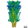
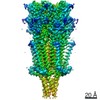
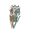
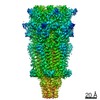
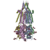
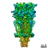
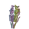
| Title | Gating movement in acetylcholine receptor analysed by time-resolved electron cryo-microscopy (closed class) | ||||||
 Components Components |
| ||||||
 Keywords Keywords | MEMBRANE PROTEIN / FREEZE-TRAPPING / ASYMMETRIC GATING / ALLOSTERIC MECHANISM | ||||||
| Function / homology |  Function and homology information Function and homology informationacetylcholine-gated channel complex / acetylcholine-gated monoatomic cation-selective channel activity / acetylcholine receptor signaling pathway / transmembrane signaling receptor activity / postsynaptic membrane Similarity search - Function | ||||||
| Biological species |  | ||||||
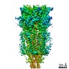
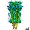
| Method | ELECTRON MICROSCOPY / helical reconstruction / cryo EM / Resolution: 6.2 Å | ||||||
 Authors Authors | Unwin, N. / Fujiyoshi, Y. | ||||||
 Citation Citation | Journal: J Mol Biol / Year: 2012 Title: Gating movement of acetylcholine receptor caught by plunge-freezing. Authors: Nigel Unwin / Yoshinori Fujiyoshi /   Abstract: The nicotinic acetylcholine (ACh) receptor converts transiently to an open-channel form when activated by ACh released into the synaptic cleft. We describe here the conformational change underlying ...The nicotinic acetylcholine (ACh) receptor converts transiently to an open-channel form when activated by ACh released into the synaptic cleft. We describe here the conformational change underlying this event, determined by electron microscopy of ACh-sprayed and freeze-trapped postsynaptic membranes. ACh binding to the α subunits triggers a concerted rearrangement in the ligand-binding domain, involving an ~1-Å outward displacement of the extracellular portion of the β subunit where it interacts with the juxtaposed ends of α-helices shaping the narrow membrane-spanning pore. The β-subunit helices tilt outward to accommodate this displacement, destabilising the arrangement of pore-lining helices, which in the closed channel bend inward symmetrically to form a central hydrophobic gate. Straightening and tangential motion of the pore-lining helices effect channel opening by widening the pore asymmetrically and increasing its polarity in the region of the gate. The pore-lining helices of the α(γ) and δ subunits, by flexing between alternative bent and straight conformations, undergo the greatest movements. This coupled allosteric transition shifts the structure from a tense (closed) state toward a more relaxed (open) state. | ||||||
| History |
|
- Structure visualization
| Movie |
Movie viewer |
|---|---|
| Structure viewer | Molecule: MolmilJmol/JSmol |
- Downloads & links
-Download
| PDBx/mmCIF format | 4aq5.cif.gz | 369.3 KB | Display | PDBx/mmCIF format |
|---|---|---|---|---|
| PDB format | pdb4aq5.ent.gz | 263.2 KB | Display | PDB format |
| PDBx/mmJSON format | 4aq5.json.gz | Tree view | PDBx/mmJSON format | |
| Others |  Other downloads Other downloads |
-Validation report
| Arichive directory | https://data.pdbj.org/pub/pdb/validation_reports/aq/4aq5ftp://data.pdbj.org/pub/pdb/validation_reports/aq/4aq5 | HTTPS FTP |
|---|
-Related structure data
| Related structure data | 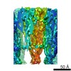 2071MC 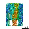 2072C  4aq9C M: map data used to model this data C: citing same article ( |
|---|---|
| Similar structure data |

-Links
 PDBj
PDBj

- Assembly
| Deposited unit | 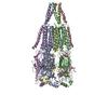
|
|---|---|
| 1 |
|
-Components
| #1: Protein | Mass: 52845.523 Da / Num. of mol.: 2 / Source method: isolated from a natural source / Details: STATION BIOLOGIQUE DE ROSCOFF / Source: (natural) #2: Protein | | Mass: 56123.594 Da / Num. of mol.: 1 / Source method: isolated from a natural source / Details: STATION BIOLOGIQUE DE ROSCOFF / Source: (natural) #3: Protein | | Mass: 60017.684 Da / Num. of mol.: 1 / Source method: isolated from a natural source / Details: STATION BIOLOGIQUE DE ROSCOFF / Source: (natural) #4: Protein | | Mass: 56234.578 Da / Num. of mol.: 1 / Source method: isolated from a natural source / Details: STATION BIOLOGIQUE DE ROSCOFF / Source: (natural) Has protein modification | Y | |
|---|
-Experimental details
-Experiment
| Experiment | Method: ELECTRON MICROSCOPY |
|---|---|
| EM experiment | Aggregation state: HELICAL ARRAY / 3D reconstruction method: helical reconstruction |
- Sample preparation
| Component | Name: NICOTINIC ACETYLCHOLINE RECEPTOR IN NATIVE POSTSYNAPTIC MEMBRANE FROM TORPEDO MARMORATA Type: COMPLEX Details: PRELIMINARY SELECTION OF MICROGRAPHS BY OPTICAL DIFFRACTIO THEN EVALUATION USING COMPUTED FOURIER TRANSFORMS |
|---|---|
| Buffer solution | Name: 100MM SODIUM CACODYLATE, 1MM CALCIUM CHLORIDE / pH: 7 / Details: 100MM SODIUM CACODYLATE, 1MM CALCIUM CHLORIDE |
| Specimen | Embedding applied: NO / Shadowing applied: NO / Staining applied: NO / Vitrification applied: YES |
| Specimen support | Details: HOLEY CARBON |
| Vitrification | Instrument: HOMEMADE PLUNGER / Cryogen name: ETHANE Details: VITRIFICATION 1 -- CRYOGEN- ETHANE, HUMIDITY- 85 TEMPERATURE- 120 INSTRUMENT- HOMEMADE PLUNGER METHOD- BLOT UNTIL APPLIED DROPLET LOSES CONTACT WITH FILTER PAPER (INDICATED BY LOSS OF ...Details: VITRIFICATION 1 -- CRYOGEN- ETHANE, HUMIDITY- 85 TEMPERATURE- 120 INSTRUMENT- HOMEMADE PLUNGER METHOD- BLOT UNTIL APPLIED DROPLET LOSES CONTACT WITH FILTER PAPER (INDICATED BY LOSS OF TRANSPARENCY TYPICALLY 6S) TIMERESOLVEDSTATE- VITRIFIED WITHIN 10MS OF EXPOSURE TO ACETYLCHOLINE (APPLIED AS THE GRID IS BEING PLUNGED USING A FINE FOCUSSED SPRAY POSITIONED ABOUT 1CM ABOVE THE ETHANE SURFACE) DETAILS- VITRIFICATION CARRIED OUT AT AN AMBIENT TEMPERATURE OF 8 DEGREES |
- Electron microscopy imaging
| Microscopy | Model: JEOL 3000SFF / Date: Nov 1, 2005 Details: STANDARD LOW DOSE IMAGING OF SPECIMENS OVER HOLES IN THE CARBON SUPPORT FILM |
|---|---|
| Electron gun | Electron source:  FIELD EMISSION GUN / Accelerating voltage: 300 kV / Illumination mode: FLOOD BEAM FIELD EMISSION GUN / Accelerating voltage: 300 kV / Illumination mode: FLOOD BEAM |
| Electron lens | Mode: BRIGHT FIELD / Nominal magnification: 40000 X / Calibrated magnification: 38500 X / Nominal defocus max: 2000 nm / Nominal defocus min: 900 nm / Cs: 1.6 mm |
| Specimen holder | Temperature: 4 K |
| Image recording | Electron dose: 25 e/Å2 / Film or detector model: KODAK SO-163 FILM |
| Image scans | Num. digital images: 111 |
| Radiation wavelength | Relative weight: 1 |
- Processing
| EM software |
| ||||||||||||
|---|---|---|---|---|---|---|---|---|---|---|---|---|---|
| CTF correction | Details: EACH TUBE IMAGE | ||||||||||||
| 3D reconstruction | Method: STANDARD FOURIER-BESSEL SYNTHESIS / Resolution: 6.2 Å / Nominal pixel size: 1 Å / Actual pixel size: 1 Å / Magnification calibration: CALIBRATION GRID IN MICROSCOPE Details: FOURIER-BESSEL SYNTHESIS AFTER APPLYING DISTORTION CORRECTIONS TO THE IMAGES. SUBMISSION BASED ON EXPERIMENTAL DATA FROM EMDB EMD-2071. Symmetry type: HELICAL | ||||||||||||
| Atomic model building | Protocol: FLEXIBLE FIT / Space: REAL Details: METHOD--MAXIMISATION OF CORRELATION BETWEEN EXPERIMENTAL DENSITIES AND ATOMIC MODEL, USING A DEFORMABLE ELASTIC NETWORK ALGORITHM (DIREX) REFINEMENT PROTOCOL--LOW RESOLUTION X-RAY | ||||||||||||
| Atomic model building | PDB-ID: 2BG9 Accession code: 2BG9 / Source name: PDB / Type: experimental model | ||||||||||||
| Refinement | Highest resolution: 6.2 Å | ||||||||||||
| Refinement step | Cycle: LAST / Highest resolution: 6.2 Å
|
