 Movie
Movie Controller

[English] 日本語
 Yorodumi
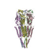
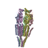
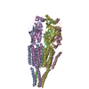
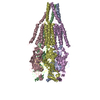
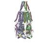
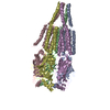
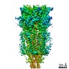
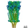
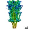
- PDB-2bg9: REFINED STRUCTURE OF THE NICOTINIC ACETYLCHOLINE RECEPTOR AT 4A R... -
+ Open data

- Basic information
| Entry | Database: PDB / ID: 2bg9 | ||||||
|---|---|---|---|---|---|---|---|
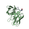
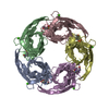
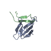
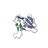
| Title | REFINED STRUCTURE OF THE NICOTINIC ACETYLCHOLINE RECEPTOR AT 4A RESOLUTION. | ||||||
 Components Components |
| ||||||
 Keywords Keywords | ION CHANNEL/RECEPTOR / ACETYLCHOLINE RECEPTOR / ION CHANNEL / ION TRANSPORT / POSTSYNAPTIC MEMBRANE / ION CHANNEL-RECEPTOR complex | ||||||
| Function / homology |  Function and homology information Function and homology informationacetylcholine-gated channel complex / acetylcholine-gated monoatomic cation-selective channel activity / acetylcholine receptor signaling pathway / transmembrane signaling receptor activity / postsynaptic membrane Similarity search - Function | ||||||
| Biological species |  | ||||||
| Method | ELECTRON MICROSCOPY / helical reconstruction / cryo EM / Resolution: 4 Å | ||||||
 Authors Authors | Unwin, N. | ||||||
 Citation Citation | Journal: J Mol Biol / Year: 2005 Title: Refined structure of the nicotinic acetylcholine receptor at 4A resolution. Authors: Nigel Unwin /  Abstract: We present a refined model of the membrane-associated Torpedo acetylcholine (ACh) receptor at 4A resolution. An improved experimental density map was obtained from 342 electron images of helical ...We present a refined model of the membrane-associated Torpedo acetylcholine (ACh) receptor at 4A resolution. An improved experimental density map was obtained from 342 electron images of helical tubes, and the refined structure was derived to an R-factor of 36.7% (R(free) 37.9%) by standard crystallographic methods, after placing the densities corresponding to a single molecule into an artificial unit cell. The agreement between experimental and calculated phases along the helical layer-lines was used to monitor progress in the refinement and to give an independent measure of the accuracy. The atomic model allowed a detailed description of the whole receptor in the closed-channel form, including the ligand-binding and intracellular domains, which have not previously been interpreted at a chemical level. We confirm that the two ligand-binding alpha subunits have a different extended conformation from the three other subunits in the closed channel, and identify several interactions on both pairs of subunit interfaces, and within the alpha subunits, which may be responsible for their "distorted" structures. The ACh-coordinating amino acid side-chains of the alpha subunits are far apart in the closed channel, indicating that a localised rearrangement, involving closure of loops B and C around the bound ACh molecule, occurs upon activation. A comparison of the structure of the alpha subunit with that of AChBP having ligand present, suggests how the localised rearrangement overcomes the distortions and initiates the rotational movements associated with opening of the channel. Both vestibules of the channel are strongly electronegative, providing a cation-stabilising environment at either entrance of the membrane pore. Access to the pore on the intracellular side is further influenced by narrow lateral windows, which would be expected to screen out electrostatically ions of the wrong charge and size. #1: Journal: Nature / Year: 2003Title: Structure and gating mechanism of the acetylcholine receptor pore. Authors: Atsuo Miyazawa / Yoshinori Fujiyoshi / Nigel Unwin /  Abstract: The nicotinic acetylcholine receptor controls electrical signalling between nerve and muscle cells by opening and closing a gated, membrane-spanning pore. Here we present an atomic model of the ...The nicotinic acetylcholine receptor controls electrical signalling between nerve and muscle cells by opening and closing a gated, membrane-spanning pore. Here we present an atomic model of the closed pore, obtained by electron microscopy of crystalline postsynaptic membranes. The pore is shaped by an inner ring of 5 alpha-helices, which curve radially to create a tapering path for the ions, and an outer ring of 15 alpha-helices, which coil around each other and shield the inner ring from the lipids. The gate is a constricting hydrophobic girdle at the middle of the lipid bilayer, formed by weak interactions between neighbouring inner helices. When acetylcholine enters the ligand-binding domain, it triggers rotations of the protein chains on opposite sides of the entrance to the pore. These rotations are communicated through the inner helices, and open the pore by breaking the girdle apart. | ||||||
| History |
| ||||||
| Remark 650 | HELIX DETERMINATION METHOD: AUTHOR PROVIDED. | ||||||
| Remark 700 | SHEET THE SHEET STRUCTURE OF THIS MOLECULE IS BIFURCATED. IN ORDER TO REPRESENT THIS FEATURE IN ... SHEET THE SHEET STRUCTURE OF THIS MOLECULE IS BIFURCATED. IN ORDER TO REPRESENT THIS FEATURE IN THE SHEET RECORDS BELOW, TWO SHEETS ARE DEFINED. |
- Structure visualization
| Movie |
Movie viewer |
|---|---|
| Structure viewer | Molecule: MolmilJmol/JSmol |
- Downloads & links
-Download
| PDBx/mmCIF format | 2bg9.cif.gz | 331.2 KB | Display | PDBx/mmCIF format |
|---|---|---|---|---|
| PDB format | pdb2bg9.ent.gz | 257.5 KB | Display | PDB format |
| PDBx/mmJSON format | 2bg9.json.gz | Tree view | PDBx/mmJSON format | |
| Others |  Other downloads Other downloads |
-Validation report
| Arichive directory | https://data.pdbj.org/pub/pdb/validation_reports/bg/2bg9ftp://data.pdbj.org/pub/pdb/validation_reports/bg/2bg9 | HTTPS FTP |
|---|
-Related structure data
| Related structure data | |
|---|---|
| Similar structure data |


-Links
 PDBj
PDBj

- Assembly
| Deposited unit | 
|
|---|---|
| 1 |
|
-Components
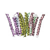
| #1: Protein | Mass: 42501.316 Da / Num. of mol.: 2 / Source method: isolated from a natural source / Source: (natural) #2: Protein | | Mass: 42167.359 Da / Num. of mol.: 1 / Source method: isolated from a natural source / Source: (natural) #3: Protein | | Mass: 42280.695 Da / Num. of mol.: 1 / Source method: isolated from a natural source / Source: (natural) #4: Protein | | Mass: 42299.496 Da / Num. of mol.: 1 / Source method: isolated from a natural source / Details: INHABITS THE EUROPEAN COAST / Source: (natural) Has protein modification | Y | |
|---|
-Experimental details
-Experiment
| Experiment | Method: ELECTRON MICROSCOPY |
|---|---|
| EM experiment | Aggregation state: HELICAL ARRAY / 3D reconstruction method: helical reconstruction |
- Sample preparation
| Component | Name: NICOTINIC ACETYLCHOLINE RECEPTOR in postsynaptic membrane Type: COMPLEX |
|---|---|
| Buffer solution | Name: 100MM SODIUM CACODYLATE / pH: 6.8 / Details: 100MM SODIUM CACODYLATE |
| Specimen | Embedding applied: NO / Shadowing applied: NO / Staining applied: NO / Vitrification applied: YES |
| Specimen support | Details: HOLEY CARBON |
| Vitrification | Details: LIQUID ETHANE |
- Electron microscopy imaging
| Microscopy | Model: JEOL 3000SFF / Date: Oct 1, 2000 Details: THE SPECIMENS WERE TUBULAR CRYSTALS GROWN IN LOW SALT BUFFER FROM ISOLATED TORPEDO POSTSYNAPTIC MEMBRANES. THEY WERE APPLIED TO THE MICROSCOPE GRIDS AND FROZEN IN THE SAME SOLUTION. THESE ...Details: THE SPECIMENS WERE TUBULAR CRYSTALS GROWN IN LOW SALT BUFFER FROM ISOLATED TORPEDO POSTSYNAPTIC MEMBRANES. THEY WERE APPLIED TO THE MICROSCOPE GRIDS AND FROZEN IN THE SAME SOLUTION. THESE CRYSTALS WERE TOO SMALL AND DISTORTED TO YIELD MEANINGFUL ELECTRON DIFFRACTION PATTERNS, SO BOTH THE AMPLITUDE AND THE PHASE TERMS HAD TO BE MEASURED FROM FOURIER TRANSFORMS OF THE IMAGES. THE IMAGES WERE RECORDED USING A DOSE OF 2000 ELECTRONS/NM2 DURING THE PERIOD: JAN-1996 TO OCT-2002. THE DATASETS INVOLVED 4 HELICAL FAMILIES OF TUBES. STRUCTURES WERE SYNTHESISED FROM THE AMPLITUDE AND PHASE TERMS DERIVED FROM EACH FAMILY. THE FINAL DATASET WAS OBTAINED BY AVERAGING THESE 4 STRUCTURES IN REAL SPACE. |
|---|---|
| Electron gun | Electron source:  FIELD EMISSION GUN / Accelerating voltage: 300 kV / Illumination mode: OTHER FIELD EMISSION GUN / Accelerating voltage: 300 kV / Illumination mode: OTHER |
| Electron lens | Mode: BRIGHT FIELD / Nominal magnification: 40000 X / Calibrated magnification: 36800 X / Nominal defocus max: 1800 nm / Nominal defocus min: 800 nm / Cs: 1.3 mm |
| Specimen holder | Temperature: 4.2 K |
| Image recording | Film or detector model: KODAK SO-163 FILM |
| Image scans | Num. digital images: 342 |
| Radiation wavelength | Relative weight: 1 |
- Processing
| 3D reconstruction | Resolution: 4 Å / Resolution method: OTHER / Symmetry type: HELICAL | ||||||||||||
|---|---|---|---|---|---|---|---|---|---|---|---|---|---|
| Refinement | Highest resolution: 4 Å | ||||||||||||
| Refinement step | Cycle: LAST / Highest resolution: 4 Å
|
