 Movie
Movie Controller

[English] 日本語
 Yorodumi
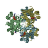
- PDB-2b8q: X-ray structure of Acanthamoeba ployphaga mimivirus nucleoside di... -
+ Open data

- Basic information
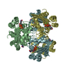
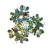
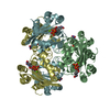
| Entry | Database: PDB / ID: 2b8q | ||||||
|---|---|---|---|---|---|---|---|
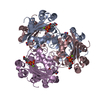
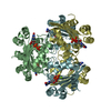
| Title | X-ray structure of Acanthamoeba ployphaga mimivirus nucleoside diphosphate kinase complexed with TDP | ||||||
 Components Components | Probable nucleoside diphosphate kinase | ||||||
 Keywords Keywords | TRANSFERASE / NDK / TDP / phosphotranferase | ||||||
| Function / homology |  Function and homology information Function and homology informationnucleoside-diphosphate kinase / UTP biosynthetic process / CTP biosynthetic process / nucleoside diphosphate kinase activity / GTP biosynthetic process / ATP binding / metal ion binding Similarity search - Function | ||||||
| Biological species |  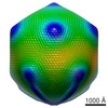 Mimivirus Mimivirus | ||||||
| Method |  X-RAY DIFFRACTION / SYNCHROTRON / MOLECULAR REPLACEMENT / Resolution: 2.5 Å X-RAY DIFFRACTION / SYNCHROTRON / MOLECULAR REPLACEMENT / Resolution: 2.5 Å | ||||||
 Authors Authors | Jeudy, S. / Claverie, J.M. / Abergel, C. | ||||||
 Citation Citation | Journal: J.Virol. / Year: 2009 Title: Dissecting the unique nucleotide specificity of mimivirus nucleoside diphosphate kinase. Authors: Jeudy, S. / Lartigue, A. / Claverie, J.M. / Abergel, C. | ||||||
| History |
|
- Structure visualization
| Structure viewer | Molecule: MolmilJmol/JSmol |
|---|
- Downloads & links
-Download
| PDBx/mmCIF format | 2b8q.cif.gz | 174.2 KB | Display | PDBx/mmCIF format |
|---|---|---|---|---|
| PDB format | pdb2b8q.ent.gz | 138.3 KB | Display | PDB format |
| PDBx/mmJSON format | 2b8q.json.gz | Tree view | PDBx/mmJSON format | |
| Others |  Other downloads Other downloads |
-Validation report
| Arichive directory | https://data.pdbj.org/pub/pdb/validation_reports/b8/2b8qftp://data.pdbj.org/pub/pdb/validation_reports/b8/2b8q | HTTPS FTP |
|---|
-Related structure data
| Related structure data | 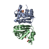 2b8pSC 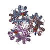 3b6bC 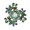 3ddiC  3dkdC 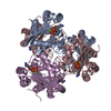 3ee3C 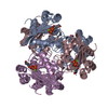 3eicC 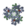 3ejmC  3elhC 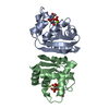 3em1C 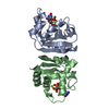 3emtC  3enaC 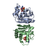 3etmC 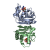 3evmC 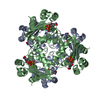 3evoC  3evwC 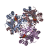 3fbbC 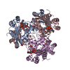 3fbcC 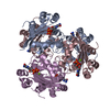 3fbeC 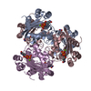 3fbfC 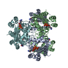 3fc9C 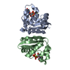 3fcvC 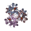 3fcwC 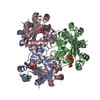 3g2xC 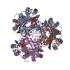 3gp9C 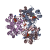 3gpaC S: Starting model for refinement C: citing same article ( |
|---|---|
| Similar structure data |


-Links
 PDBj
PDBj- Assembly
| Deposited unit | 
| ||||||||
|---|---|---|---|---|---|---|---|---|---|
| 1 | 
| ||||||||
| 2 | 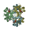
| ||||||||
| Unit cell |
|
-Components
| #1: Protein | Mass: 16301.495 Da / Num. of mol.: 6 Source method: isolated from a genetically manipulated source Source: (gene. exp.) Mimivirus / Genus: Mimivirus / Gene: NDK / Plasmid: pDIGS02 / Production host:  #2: Chemical | ChemComp-MG /   Mass: 24.305 Da / Num. of mol.: 6 / Source method: obtained synthetically / Formula: Mg Mass: 24.305 Da / Num. of mol.: 6 / Source method: obtained synthetically / Formula: Mg#3: Chemical | ChemComp-TYD /   Mass: 402.188 Da / Num. of mol.: 6 / Source method: obtained synthetically / Formula: C10H16N2O11P2 Mass: 402.188 Da / Num. of mol.: 6 / Source method: obtained synthetically / Formula: C10H16N2O11P2#4: Chemical | ChemComp-PO4 / |   Mass: 94.971 Da / Num. of mol.: 1 / Source method: obtained synthetically / Formula: PO4 Mass: 94.971 Da / Num. of mol.: 1 / Source method: obtained synthetically / Formula: PO4#5: Water | ChemComp-HOH / |  Mass: 18.015 Da / Num. of mol.: 153 / Source method: isolated from a natural source / Formula: H2O Mass: 18.015 Da / Num. of mol.: 153 / Source method: isolated from a natural source / Formula: H2O |
|---|
-Experimental details
-Experiment
| Experiment | Method: X-RAY DIFFRACTION / Number of used crystals: 1 |
|---|
- Sample preparation
| Crystal | Density Matthews: 3.14 Å3/Da / Density % sol: 60.5 % |
|---|---|
| Crystal grow | Temperature: 293 K / Method: vapor diffusion, hanging drop / pH: 7.5 Details: 44% MPD, 0.1M hepes, pH 7.5, VAPOR DIFFUSION, HANGING DROP, temperature 293K |
-Data collection
| Diffraction | Mean temperature: 100 K |
|---|---|
| Diffraction source | Source: SYNCHROTRON / Site: ESRF  / Beamline: ID23-1 / Wavelength: 0.97565 Å / Beamline: ID23-1 / Wavelength: 0.97565 Å |
| Detector | Type: MARRESEARCH / Detector: CCD / Date: Jul 6, 2005 / Details: mirrors |
| Radiation | Monochromator: silicon / Protocol: SINGLE WAVELENGTH / Monochromatic (M) / Laue (L): M / Scattering type: x-ray |
| Radiation wavelength | Wavelength: 0.97565 Å / Relative weight: 1 |
| Reflection | Resolution: 2.5→66.5 Å / Num. obs: 39727 / % possible obs: 100 % / Observed criterion σ(F): 0 / Observed criterion σ(I): 0 / Redundancy: 5 % / Biso Wilson estimate: 51 Å2 / Rmerge(I) obs: 0.079 / Rsym value: 0.079 / Net I/σ(I): 5.9 |
| Reflection shell | Resolution: 2.5→2.64 Å / Redundancy: 5 % / Rmerge(I) obs: 0.355 / Mean I/σ(I) obs: 2 / Rsym value: 0.355 / % possible all: 100 |
- Processing
| Software |
| ||||||||||||||||||||||||||||||||||||||||||||||||||||||||||||
|---|---|---|---|---|---|---|---|---|---|---|---|---|---|---|---|---|---|---|---|---|---|---|---|---|---|---|---|---|---|---|---|---|---|---|---|---|---|---|---|---|---|---|---|---|---|---|---|---|---|---|---|---|---|---|---|---|---|---|---|---|---|
| Refinement | Method to determine structure: MOLECULAR REPLACEMENT Starting model: PDB ENTRY 2B8P Resolution: 2.5→29.55 Å / Rfactor Rfree error: 0.005 / Data cutoff high absF: 2483223.13 / Data cutoff low absF: 0 / Isotropic thermal model: RESTRAINED / Cross valid method: THROUGHOUT / σ(F): 0 / Details: REFMAC was also used for the refinement.
| ||||||||||||||||||||||||||||||||||||||||||||||||||||||||||||
| Solvent computation | Solvent model: FLAT MODEL / Bsol: 31.6874 Å2 / ksol: 0.324969 e/Å3 | ||||||||||||||||||||||||||||||||||||||||||||||||||||||||||||
| Displacement parameters | Biso mean: 44.6 Å2
| ||||||||||||||||||||||||||||||||||||||||||||||||||||||||||||
| Refine analyze |
| ||||||||||||||||||||||||||||||||||||||||||||||||||||||||||||
| Refinement step | Cycle: LAST / Resolution: 2.5→29.55 Å
| ||||||||||||||||||||||||||||||||||||||||||||||||||||||||||||
| Refine LS restraints |
| ||||||||||||||||||||||||||||||||||||||||||||||||||||||||||||
| LS refinement shell | Resolution: 2.5→2.66 Å / Rfactor Rfree error: 0.019 / Total num. of bins used: 6
| ||||||||||||||||||||||||||||||||||||||||||||||||||||||||||||
| Xplor file |
|
