 Movie
Movie Controller

[English] 日本語
 Yorodumi
- PDB-2axq: Apo histidine-tagged saccharopine dehydrogenase (L-Glu forming) f... -
+ Open data

- Basic information
| Entry | Database: PDB / ID: 2axq | ||||||
|---|---|---|---|---|---|---|---|
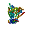
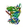
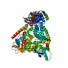
| Title | Apo histidine-tagged saccharopine dehydrogenase (L-Glu forming) from Saccharomyces cerevisiae | ||||||
 Components Components | Saccharopine dehydrogenase | ||||||
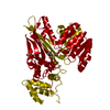
 Keywords Keywords | OXIDOREDUCTASE / ROSSMANN FOLD VARIANT / SACCHAROPINE REDUCTASE FOLD (DOMAIN II) / ALPHA/BETA PROTEIN / ALPHA-AMINOADIPATE PATHWAY / FUNGAL LYSINE BIOSYNTHESIS | ||||||
| Function / homology |  Function and homology information Function and homology informationLysine catabolism / saccharopine dehydrogenase (NADP+, L-glutamate-forming) / saccharopine dehydrogenase (NADP+, L-glutamate-forming) activity / saccharopine dehydrogenase activity / lysine biosynthetic process via aminoadipic acid / cell periphery / cytoplasm Similarity search - Function | ||||||
| Biological species |  | ||||||
| Method |  X-RAY DIFFRACTION / MOLECULAR REPLACEMENT / Resolution: 1.7 Å X-RAY DIFFRACTION / MOLECULAR REPLACEMENT / Resolution: 1.7 Å | ||||||
 Authors Authors | Andi, B. / Cook, P.F. / West, A.H. | ||||||
 Citation Citation | Journal: Cell Biochem.Biophys. / Year: 2006 Title: Crystal structure of the his-tagged saccharopine reductase from Saccharomyces cerevisiae at 1.7-A resolution. Authors: Andi, B. / Cook, P.F. / West, A.H. #1: Journal: Structure Fold.Des. / Year: 2000Title: Crystal Structure of Saccharopine Reductase from Magnaporthe Grisea, an Enzyme of the Alpha-Aminoadipate Pathway of Lysine Biosynthesis Authors: Johansson, E. / Steffens, J.J. / Lindqvist, Y. / Schneider, G. | ||||||
| History |
|
- Structure visualization
| Structure viewer | Molecule: MolmilJmol/JSmol |
|---|
- Downloads & links
-Download
| PDBx/mmCIF format | 2axq.cif.gz | 109.7 KB | Display | PDBx/mmCIF format |
|---|---|---|---|---|
| PDB format | pdb2axq.ent.gz | 81.9 KB | Display | PDB format |
| PDBx/mmJSON format | 2axq.json.gz | Tree view | PDBx/mmJSON format | |
| Others |  Other downloads Other downloads |
-Validation report
| Summary document | 2axq_validation.pdf.gz | 440.4 KB | Display | wwPDB validaton report |
|---|---|---|---|---|
| Full document | 2axq_full_validation.pdf.gz | 441.3 KB | Display | |
| Data in XML | 2axq_validation.xml.gz | 20.8 KB | Display | |
| Data in CIF | 2axq_validation.cif.gz | 31.3 KB | Display | |
| Arichive directory | https://data.pdbj.org/pub/pdb/validation_reports/ax/2axqftp://data.pdbj.org/pub/pdb/validation_reports/ax/2axq | HTTPS FTP |
-Related structure data
| Related structure data |  1e5lS S: Starting model for refinement |
|---|---|
| Similar structure data |


-Links
 PDBj
PDBj- Assembly
| Deposited unit | 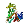
| ||||||||
|---|---|---|---|---|---|---|---|---|---|
| 1 | 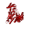
| ||||||||
| Unit cell |
| ||||||||
| Components on special symmetry positions |
| ||||||||
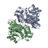
| Details | The biological assembly is a homodimer generated from the monomer in the asymmetric unit by the operations: y, x, -z |
-Components
| #1: Protein | Mass: 51505.371 Da / Num. of mol.: 1 Source method: isolated from a genetically manipulated source Details: L-Glu forming Source: (gene. exp.) Gene: LYS9, LYS13 / Plasmid: pET-16b-LYS9 / Production host:  References: UniProt: P38999, saccharopine dehydrogenase (NADP+, L-glutamate-forming) | ||
|---|---|---|---|
| #2: Chemical | ChemComp-SO4 /   Mass: 96.063 Da / Num. of mol.: 4 / Source method: obtained synthetically / Formula: SO4 Mass: 96.063 Da / Num. of mol.: 4 / Source method: obtained synthetically / Formula: SO4#3: Water | ChemComp-HOH / |  Mass: 18.015 Da / Num. of mol.: 349 / Source method: isolated from a natural source / Formula: H2O Mass: 18.015 Da / Num. of mol.: 349 / Source method: isolated from a natural source / Formula: H2O |
-Experimental details
-Experiment
| Experiment | Method: X-RAY DIFFRACTION / Number of used crystals: 1 |
|---|
- Sample preparation
| Crystal | Density Matthews: 2.9 Å3/Da / Density % sol: 57.5 % |
|---|---|
| Crystal grow | Temperature: 298 K / Method: vapor diffusion, hanging drop / pH: 7.2 Details: 1.2M Ammonium sulfate, 25mM Bis-Tris, 6.5-7mg/mL enzyme, 50mM Tris-HCl pH 8.0, 150mM KCl, 75mM imidazole, pH 7.2, VAPOR DIFFUSION, HANGING DROP, temperature 298.0K |
-Data collection
| Diffraction | Mean temperature: 100 K |
|---|---|
| Diffraction source | Source: ROTATING ANODE / Type: RIGAKU RUH3R / Wavelength: 1.5418 Å |
| Detector | Type: RIGAKU RAXIS IV / Detector: IMAGE PLATE / Date: Aug 18, 2003 / Details: MICRO-OPTICS |
| Radiation | Monochromator: OSMIC CONFOCAL OPTICS / Protocol: SINGLE WAVELENGTH / Monochromatic (M) / Laue (L): M / Scattering type: x-ray |
| Radiation wavelength | Wavelength: 1.5418 Å / Relative weight: 1 |
| Reflection | Resolution: 1.7→39.85 Å / Num. all: 66370 / Num. obs: 64775 / % possible obs: 97.6 % / Observed criterion σ(F): 0 / Observed criterion σ(I): 2 / Redundancy: 7.18 % / Biso Wilson estimate: 23.6 Å2 / Rmerge(I) obs: 0.056 / Χ2: 1 / Net I/σ(I): 15.8 / Scaling rejects: 3516 |
| Reflection shell | Resolution: 1.7→1.76 Å / % possible obs: 95.9 % / Redundancy: 4.98 % / Rmerge(I) obs: 0.368 / Mean I/σ(I) obs: 4.3 / Num. measured obs: 324 / Num. unique all: 6559 / Χ2: 1.16 / % possible all: 95.9 |
-Phasing
| Phasing MR |
|
|---|
- Processing
| Software |
| ||||||||||||||||||||||||||||||||||||||||||||||||||||||||||||||||||||||||||||||||||||||||||
|---|---|---|---|---|---|---|---|---|---|---|---|---|---|---|---|---|---|---|---|---|---|---|---|---|---|---|---|---|---|---|---|---|---|---|---|---|---|---|---|---|---|---|---|---|---|---|---|---|---|---|---|---|---|---|---|---|---|---|---|---|---|---|---|---|---|---|---|---|---|---|---|---|---|---|---|---|---|---|---|---|---|---|---|---|---|---|---|---|---|---|---|
| Refinement | Method to determine structure: MOLECULAR REPLACEMENT Starting model: PDB ENTRY 1E5L, used monomer, backbone only Resolution: 1.7→39.85 Å / Cor.coef. Fo:Fc: 0.962 / Cor.coef. Fo:Fc free: 0.942 / SU B: 2.661 / SU ML: 0.084 / Isotropic thermal model: Mixed (isotropic/anisotropic) / Cross valid method: THROUGHOUT / σ(F): 0 / σ(I): 2 / ESU R: 0.099 / ESU R Free: 0.104 / Stereochemistry target values: MAXIMUM LIKELIHOOD Details: The first methionine residue and N-terminal deca-histidine tag could not be built because of missing electron density.
| ||||||||||||||||||||||||||||||||||||||||||||||||||||||||||||||||||||||||||||||||||||||||||
| Solvent computation | Ion probe radii: 0.8 Å / Shrinkage radii: 0.8 Å / VDW probe radii: 1.2 Å / Solvent model: BABINET MODEL WITH MASK | ||||||||||||||||||||||||||||||||||||||||||||||||||||||||||||||||||||||||||||||||||||||||||
| Displacement parameters | Biso mean: 27.461 Å2
| ||||||||||||||||||||||||||||||||||||||||||||||||||||||||||||||||||||||||||||||||||||||||||
| Refine analyze | Luzzati coordinate error obs: 0.219 Å | ||||||||||||||||||||||||||||||||||||||||||||||||||||||||||||||||||||||||||||||||||||||||||
| Refinement step | Cycle: LAST / Resolution: 1.7→39.85 Å
| ||||||||||||||||||||||||||||||||||||||||||||||||||||||||||||||||||||||||||||||||||||||||||
| Refine LS restraints |
| ||||||||||||||||||||||||||||||||||||||||||||||||||||||||||||||||||||||||||||||||||||||||||
| LS refinement shell | Resolution: 1.7→1.744 Å / Total num. of bins used: 20
|
