[English] 日本語
Yorodumi
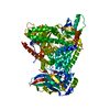
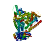
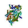
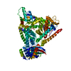
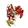
- PDB-5o1n: Crystal structure of human aminoadipate semialdehyde synthase, sa... -

+
Open data


ID or keywords:

Loading...

-
Basic information

Entry
Database: PDB / ID: 5o1n
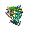
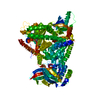
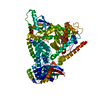
TitleCrystal structure of human aminoadipate semialdehyde synthase, saccharopine dehydrogenase domain with N-[(2S)-2-Pyrrolidinylmethyl]-trifluoromethanesulfonamide bound
ComponentsAlpha-aminoadipic semialdehyde synthase, mitochondrial
KeywordsOXIDOREDUCTASE / human aminoadipate semialdehyde synthase / saccharopine dehydrogenase domain with N-[(2S)-2-Pyrrolidinylmethyl]-trifluoromethanesulfonamide bound
Function / homology
Function and homology information


saccharopine dehydrogenase (NADP+, L-lysine-forming) / saccharopine dehydrogenase (NAD+, L-glutamate-forming) / saccharopine dehydrogenase (NADP+, L-lysine-forming) activity / saccharopine dehydrogenase (NAD+, L-glutamate-forming) activity / saccharopine dehydrogenase activity / saccharopine dehydrogenase (NAD+, L-lysine-forming) activity / lysine catabolic process / L-lysine catabolic process to acetyl-CoA via saccharopine / lysine biosynthetic process via aminoadipic acid / Lysine catabolism ...saccharopine dehydrogenase (NADP+, L-lysine-forming) / saccharopine dehydrogenase (NAD+, L-glutamate-forming) / saccharopine dehydrogenase (NADP+, L-lysine-forming) activity / saccharopine dehydrogenase (NAD+, L-glutamate-forming) activity / saccharopine dehydrogenase activity / saccharopine dehydrogenase (NAD+, L-lysine-forming) activity / lysine catabolic process / L-lysine catabolic process to acetyl-CoA via saccharopine / lysine biosynthetic process via aminoadipic acid / Lysine catabolism / transcription corepressor activity / histone binding / mitochondrial matrix / intracellular membrane-bounded organelle / negative regulation of transcription by RNA polymerase II / mitochondrion / nucleus / cytoplasm / cytosol
Similarity search - Function
Saccharopine dehydrogenase, NADP binding domain / Saccharopine dehydrogenase-like, C-terminal / Saccharopine dehydrogenase NADP binding domain / Saccharopine dehydrogenase C-terminal domain / Alanine dehydrogenase/pyridine nucleotide transhydrogenase, N-terminal / Alanine dehydrogenase/PNT, N-terminal domain / Alanine dehydrogenase/PNT, C-terminal domain / Alanine dehydrogenase/PNT, N-terminal domain / Alanine dehydrogenase/pyridine nucleotide transhydrogenase, NAD(H)-binding domain / Dihydrodipicolinate Reductase; domain 2 ...Saccharopine dehydrogenase, NADP binding domain / Saccharopine dehydrogenase-like, C-terminal / Saccharopine dehydrogenase NADP binding domain / Saccharopine dehydrogenase C-terminal domain / Alanine dehydrogenase/pyridine nucleotide transhydrogenase, N-terminal / Alanine dehydrogenase/PNT, N-terminal domain / Alanine dehydrogenase/PNT, C-terminal domain / Alanine dehydrogenase/PNT, N-terminal domain / Alanine dehydrogenase/pyridine nucleotide transhydrogenase, NAD(H)-binding domain / Dihydrodipicolinate Reductase; domain 2 / Dihydrodipicolinate Reductase; domain 2 / NAD(P)-binding Rossmann-like Domain / NAD(P)-binding domain superfamily / Rossmann fold / 2-Layer Sandwich / 3-Layer(aba) Sandwich / Alpha Beta
Similarity search - Domain/homology
Chem-9H8 / DI(HYDROXYETHYL)ETHER / Alpha-aminoadipic semialdehyde synthase, mitochondrial
Similarity search - Component
Biological speciesHomo sapiens (human)
MethodX-RAY DIFFRACTION / SYNCHROTRON / MOLECULAR REPLACEMENT / Resolution: 2.28 Å
AuthorsKopec, J. / Rembeza, E. / Pena, I.A. / Strain-Damerell, C. / Goubin, S. / Sethi, R. / Velupillai, S. / Talon, R. / Collins, P. / Krojer, T. ...Kopec, J. / Rembeza, E. / Pena, I.A. / Strain-Damerell, C. / Goubin, S. / Sethi, R. / Velupillai, S. / Talon, R. / Collins, P. / Krojer, T. / McLaughlin, M. / Burgess-Brown, N. / Arrowsmith, C. / Edwards, A. / Bountra, C. / Brennan, P. / von Delft, F. / Arruda, P. / Yue, W.W.
CitationJournal: To Be Published
Title: Crystal structure of human aminoadipate semialdehyde synthase, saccharopine dehydrogenase domain with N-[(2S)-2-Pyrrolidinylmethyl]-trifluoromethanesulfonamide bound
Authors: Kopec, J. / Yue, W.W.
History
DepositionMay 18, 2017Deposition site: PDBE / Processing site: PDBE
Revision 1.0May 31, 2017Provider: repository / Type: Initial release
Revision 1.1Jan 17, 2024Group: Data collection / Database references / Refinement description
Category: chem_comp_atom / chem_comp_bond ...chem_comp_atom / chem_comp_bond / database_2 / pdbx_initial_refinement_model
Item: _database_2.pdbx_DOI / _database_2.pdbx_database_accession

-
Structure visualization

Structure viewerMolecule:
MolmilJmol/JSmol

Downloads & links

-
Assembly

Deposited unit
A: Alpha-aminoadipic semialdehyde synthase, mitochondrial
hetero molecules


Theoretical massNumber of molelcules
Total (without water)55,0875
Polymers54,6091
Non-polymers4794
Water45025
1
A: Alpha-aminoadipic semialdehyde synthase, mitochondrial
hetero molecules

A: Alpha-aminoadipic semialdehyde synthase, mitochondrial
hetero molecules


Theoretical massNumber of molelcules
Total (without water)110,17510
Polymers109,2182
Non-polymers9578
Water362
TypeNameSymmetry operationNumber
identity operation1_555x,y,z1
crystal symmetry operation7_467y-1,x+1,-z+21
Buried area3400 Å2
ΔGint7 kcal/mol
Surface area35860 Å2
MethodPISA
Unit cell
Length a, b, c (Å)89.396, 89.396, 147.893
Angle α, β, γ (deg.)90.00, 90.00, 90.00
Int Tables number96
Space group name H-MP43212

-
Components

-
Protein , 1 types, 1 molecules A

#1: Protein Alpha-aminoadipic semialdehyde synthase, mitochondrial / LKR/SDH


Mass: 54608.812 Da / Num. of mol.: 1 / Fragment: UNP residues 455-926
Source method: isolated from a genetically manipulated source
Source: (gene. exp.) Homo sapiens (human) / Gene: AASS / Plasmid: pFB-LIC-Bse / Cell line (production host): SF9 / Production host: 
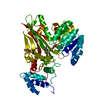
Spodoptera frugiperda (fall armyworm)
References: UniProt: Q9UDR5, saccharopine dehydrogenase (NADP+, L-lysine-forming), saccharopine dehydrogenase (NAD+, L-glutamate-forming)

-
Non-polymers , 5 types, 29 molecules

#2: Chemical ChemComp-9H8 / 1,1,1-tris(fluoranyl)-~{N}-[[(2~{S})-pyrrolidin-2-yl]methyl]methanesulfonamide


Mass: 232.224 Da / Num. of mol.: 1 / Source method: obtained synthetically / Formula: C6H11F3N2O2S
#3: Chemical ChemComp-DMS / DIMETHYL SULFOXIDE


Mass: 78.133 Da / Num. of mol.: 1 / Source method: obtained synthetically / Formula: C2H6OS / Comment: DMSO, precipitant*YM
#4: Chemical ChemComp-EDO / 1,2-ETHANEDIOL / ETHYLENE GLYCOL


Mass: 62.068 Da / Num. of mol.: 1 / Source method: obtained synthetically / Formula: C2H6O2
#5: Chemical ChemComp-PEG / DI(HYDROXYETHYL)ETHER


Mass: 106.120 Da / Num. of mol.: 1 / Source method: obtained synthetically / Formula: C4H10O3
#6: Water ChemComp-HOH / water


Mass: 18.015 Da / Num. of mol.: 25 / Source method: isolated from a natural source / Formula: H2O

-
Experimental details

-
Experiment

ExperimentMethod: X-RAY DIFFRACTION / Number of used crystals: 1

-
Sample preparation

CrystalDensity Matthews: 3.22 Å3/Da / Density % sol: 61.79 %
Crystal growTemperature: 285 K / Method: vapor diffusion, hanging drop / pH: 7.5
Details: 25% PEG3350 -- 0.3M sodium malonate -- 0.1M tris pH 7.5

-
Data collection

DiffractionMean temperature: 100 K
Diffraction sourceSource: SYNCHROTRON / Site: Diamond / Beamline: I03 / Wavelength: 0.97623 Å
DetectorType: DECTRIS PILATUS3 S 6M / Detector: PIXEL / Date: May 5, 2017
RadiationProtocol: SINGLE WAVELENGTH / Monochromatic (M) / Laue (L): M / Scattering type: x-ray
Radiation wavelengthWavelength: 0.97623 Å / Relative weight: 1
ReflectionResolution: 2.28→44.7 Å / Num. obs: 28144 / % possible obs: 100 % / Redundancy: 13.3 % / Biso Wilson estimate: 55.5 Å2 / CC1/2: 1 / Rmerge(I) obs: 0.076 / Net I/σ(I): 15.9
Reflection shellResolution: 2.28→2.34 Å / Redundancy: 10.8 % / Mean I/σ(I) obs: 1.3 / Num. unique obs: 2038 / CC1/2: 0.758 / % possible all: 100

-
Processing

Software
NameVersionClassification
REFMAC5.8.0135refinement
xia2data reduction
xia2data scaling
PHASERphasing
RefinementMethod to determine structure: MOLECULAR REPLACEMENT
Starting model: 5l76
Resolution: 2.28→44.7 Å / Cor.coef. Fo:Fc: 0.942 / Cor.coef. Fo:Fc free: 0.933 / SU B: 12.463 / SU ML: 0.27 / Cross valid method: THROUGHOUT / ESU R: 0.311 / ESU R Free: 0.236 / Details: HYDROGENS HAVE BEEN ADDED IN THE RIDING POSITIONS
RfactorNum. reflection% reflectionSelection details
Rfree0.28559 1371 4.9 %RANDOM
Rwork0.26483 ---
obs0.26585 26469 99.11 %-
Solvent computationIon probe radii: 0.8 Å / Shrinkage radii: 0.8 Å / VDW probe radii: 1.2 Å
Displacement parametersBiso mean: 68.328 Å2
Baniso -1Baniso -2Baniso -3
1-2.58 Å2-0 Å2-0 Å2
2--2.58 Å2-0 Å2
3----5.16 Å2
Refinement stepCycle: 1 / Resolution: 2.28→44.7 Å
ProteinNucleic acidLigandSolventTotal
Num. atoms3284 0 29 25 3338
Refine LS restraints
Refine-IDTypeDev idealDev ideal targetNumber
X-RAY DIFFRACTIONr_bond_refined_d0.0070.0193373
X-RAY DIFFRACTIONr_bond_other_d0.0010.023257
X-RAY DIFFRACTIONr_angle_refined_deg0.9971.994577
X-RAY DIFFRACTIONr_angle_other_deg0.8352.9957504
X-RAY DIFFRACTIONr_dihedral_angle_1_deg4.8335440
X-RAY DIFFRACTIONr_dihedral_angle_2_deg35.17625.169118
X-RAY DIFFRACTIONr_dihedral_angle_3_deg11.16815557
X-RAY DIFFRACTIONr_dihedral_angle_4_deg11.686159
X-RAY DIFFRACTIONr_chiral_restr0.0550.2537
X-RAY DIFFRACTIONr_gen_planes_refined0.0040.0213764
X-RAY DIFFRACTIONr_gen_planes_other0.0010.02675
X-RAY DIFFRACTIONr_nbd_refined
X-RAY DIFFRACTIONr_nbd_other
X-RAY DIFFRACTIONr_nbtor_refined
X-RAY DIFFRACTIONr_nbtor_other
X-RAY DIFFRACTIONr_xyhbond_nbd_refined
X-RAY DIFFRACTIONr_xyhbond_nbd_other
X-RAY DIFFRACTIONr_metal_ion_refined
X-RAY DIFFRACTIONr_metal_ion_other
X-RAY DIFFRACTIONr_symmetry_vdw_refined
X-RAY DIFFRACTIONr_symmetry_vdw_other
X-RAY DIFFRACTIONr_symmetry_hbond_refined
X-RAY DIFFRACTIONr_symmetry_hbond_other
X-RAY DIFFRACTIONr_symmetry_metal_ion_refined
X-RAY DIFFRACTIONr_symmetry_metal_ion_other
X-RAY DIFFRACTIONr_mcbond_it1.2257.0441766
X-RAY DIFFRACTIONr_mcbond_other1.2257.0411765
X-RAY DIFFRACTIONr_mcangle_it2.20910.5582204
X-RAY DIFFRACTIONr_mcangle_other2.20910.562205
X-RAY DIFFRACTIONr_scbond_it0.9386.9821606
X-RAY DIFFRACTIONr_scbond_other0.9356.9821606
X-RAY DIFFRACTIONr_scangle_it
X-RAY DIFFRACTIONr_scangle_other1.68410.4682374
X-RAY DIFFRACTIONr_long_range_B_refined3.49254.3033522
X-RAY DIFFRACTIONr_long_range_B_other3.49254.3133523
X-RAY DIFFRACTIONr_rigid_bond_restr
X-RAY DIFFRACTIONr_sphericity_free
X-RAY DIFFRACTIONr_sphericity_bonded
LS refinement shellResolution: 2.28→2.339 Å / Total num. of bins used: 20
RfactorNum. reflection% reflection
Rfree0.466 97 -
Rwork0.45 1887 -
obs--97.78 %

+
About Yorodumi

-
News

-
Feb 9, 2022. New format data for meta-information of EMDB entries

New format data for meta-information of EMDB entries

  • Version 3 of the EMDB header file is now the official format.
  • The previous official version 1.9 will be removed from the archive.

Related info.:EMDB header

External links:wwPDB to switch to version 3 of the EMDB data model

-
Aug 12, 2020. Covid-19 info

Covid-19 info

URL: https://pdbjlvh1.pdbj.org/emnavi/covid19.php

New page: Covid-19 featured information page in EM Navigator.

Related info.:Covid-19 info / Mar 5, 2020. Novel coronavirus structure data

+
Mar 5, 2020. Novel coronavirus structure data

Novel coronavirus structure data

Related info.:Yorodumi Speices / Aug 12, 2020. Covid-19 info

External links:COVID-19 featured content - PDBj / Molecule of the Month (242):Coronavirus Proteases

+
Jan 31, 2019. EMDB accession codes are about to change! (news from PDBe EMDB page)

EMDB accession codes are about to change! (news from PDBe EMDB page)

  • The allocation of 4 digits for EMDB accession codes will soon come to an end. Whilst these codes will remain in use, new EMDB accession codes will include an additional digit and will expand incrementally as the available range of codes is exhausted. The current 4-digit format prefixed with “EMD-” (i.e. EMD-XXXX) will advance to a 5-digit format (i.e. EMD-XXXXX), and so on. It is currently estimated that the 4-digit codes will be depleted around Spring 2019, at which point the 5-digit format will come into force.
  • The EM Navigator/Yorodumi systems omit the EMD- prefix.

Related info.:Q: What is EMD? / ID/Accession-code notation in Yorodumi/EM Navigator

External links:EMDB Accession Codes are Changing Soon! / Contact to PDBj

+
Jul 12, 2017. Major update of PDB

Major update of PDB

  • wwPDB released updated PDB data conforming to the new PDBx/mmCIF dictionary.
  • This is a major update changing the version number from 4 to 5, and with Remediation, in which all the entries are updated.
  • In this update, many items about electron microscopy experimental information are reorganized (e.g. em_software).
  • Now, EM Navigator and Yorodumi are based on the updated data.

External links:wwPDB Remediation / Enriched Model Files Conforming to OneDep Data Standards Now Available in the PDB FTP Archive

-
Yorodumi

Thousand views of thousand structures

  • Yorodumi is a browser for structure data from EMDB, PDB, SASBDB, etc.
  • This page is also the successor to EM Navigator detail page, and also detail information page/front-end page for Omokage search.
  • The word "yorodu" (or yorozu) is an old Japanese word meaning "ten thousand". "mi" (miru) is to see.

Related info.:EMDB / PDB / SASBDB / Comparison of 3 databanks / Yorodumi Search / Aug 31, 2016. New EM Navigator & Yorodumi / Yorodumi Papers / Jmol/JSmol / Function and homology information / Changes in new EM Navigator and Yorodumi

Read more