 Movie
Movie Controller

[English] 日本語
 Yorodumi
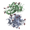
- PDB-1e5q: Ternary complex of saccharopine reductase from Magnaporthe grisea... -
+ Open data

- Basic information
| Entry | Database: PDB / ID: 1e5q | ||||||
|---|---|---|---|---|---|---|---|
| Title | Ternary complex of saccharopine reductase from Magnaporthe grisea, NADPH and saccharopine | ||||||
 Components Components | Saccharopine dehydrogenase [NADP(+), L-glutamate-forming] | ||||||
 Keywords Keywords | OXIDOREDUCTASE / SACCHAROPINE REDUCTASE / NADPH / LYSINE BIOSYNTHESIS / ALPHA- AMINOADIPATE PATHWAY | ||||||
| Function / homology |  Function and homology information Function and homology informationsaccharopine dehydrogenase (NADP+, L-glutamate-forming) / saccharopine dehydrogenase (NADP+, L-glutamate-forming) activity / : / cytoplasm Similarity search - Function | ||||||
| Biological species |  Magnaporthe oryzae (rice blast fungus) Magnaporthe oryzae (rice blast fungus) | ||||||
| Method |  X-RAY DIFFRACTION / SYNCHROTRON / MOLECULAR REPLACEMENT / Resolution: 2.1 Å X-RAY DIFFRACTION / SYNCHROTRON / MOLECULAR REPLACEMENT / Resolution: 2.1 Å | ||||||
 Authors Authors | Johansson, E. / Steffens, J.J. / Lindqvist, Y. / Schneider, G. | ||||||
 Citation Citation | Journal: Structure / Year: 2000 Title: Crystal Structure of Saccharopine Reductase from Magnaporthe Grisea, an Enzyme of the Alpha-Aminoadipate Pathway of Lysine Biosynthesis Authors: Johansson, E. / Steffens, J.J. / Lindqvist, Y. / Schneider, G. | ||||||
| History |
|
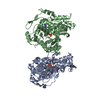
- Structure visualization
| Structure viewer | Molecule: MolmilJmol/JSmol |
|---|
- Downloads & links
-Download
| PDBx/mmCIF format | 1e5q.cif.gz | 730 KB | Display | PDBx/mmCIF format |
|---|---|---|---|---|
| PDB format | pdb1e5q.ent.gz | 607.1 KB | Display | PDB format |
| PDBx/mmJSON format | 1e5q.json.gz | Tree view | PDBx/mmJSON format | |
| Others |  Other downloads Other downloads |
-Validation report
| Arichive directory | https://data.pdbj.org/pub/pdb/validation_reports/e5/1e5qftp://data.pdbj.org/pub/pdb/validation_reports/e5/1e5q | HTTPS FTP |
|---|
-Related structure data
| Related structure data |  1e5lC  1ff9SC C: citing same article ( S: Starting model for refinement |
|---|---|
| Similar structure data |


-Links
 PDBj
PDBj

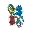
- Assembly
| Deposited unit | 
| ||||||||||||||||||||||||||||||||
|---|---|---|---|---|---|---|---|---|---|---|---|---|---|---|---|---|---|---|---|---|---|---|---|---|---|---|---|---|---|---|---|---|---|
| 1 | 
| ||||||||||||||||||||||||||||||||
| 2 | 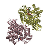
| ||||||||||||||||||||||||||||||||
| 3 | 
| ||||||||||||||||||||||||||||||||
| 4 | 
| ||||||||||||||||||||||||||||||||
| Unit cell |
| ||||||||||||||||||||||||||||||||
| Noncrystallographic symmetry (NCS) | NCS oper:
|
-Components
| #1: Protein | Mass: 49156.262 Da / Num. of mol.: 8 Source method: isolated from a genetically manipulated source Source: (gene. exp.) Magnaporthe oryzae (rice blast fungus) / Gene: LYS3, MGG_08564 / Plasmid: PDB45 / Cellular location (production host): CYTOPLASM / Production host:  References: UniProt: Q9P4R4, saccharopine dehydrogenase (NADP+, L-glutamate-forming) #2: Chemical | ChemComp-NDP /   Mass: 745.421 Da / Num. of mol.: 8 / Source method: obtained synthetically / Formula: C21H30N7O17P3 Mass: 745.421 Da / Num. of mol.: 8 / Source method: obtained synthetically / Formula: C21H30N7O17P3#3: Chemical | ChemComp-SHR /   Type: L-peptide linking / Mass: 276.286 Da / Num. of mol.: 8 / Source method: obtained synthetically / Formula: C11H20N2O6 Type: L-peptide linking / Mass: 276.286 Da / Num. of mol.: 8 / Source method: obtained synthetically / Formula: C11H20N2O6#4: Water | ChemComp-HOH / |  Mass: 18.015 Da / Num. of mol.: 2509 / Source method: isolated from a natural source / Formula: H2O Mass: 18.015 Da / Num. of mol.: 2509 / Source method: isolated from a natural source / Formula: H2OSequence details | DESCRIBED IN JOHANSSON ET AL.(2000) ACTA CRYST. D56 662-664 | |
|---|
-Experimental details
-Experiment
| Experiment | Method: X-RAY DIFFRACTION / Number of used crystals: 1 |
|---|
- Sample preparation
| Crystal | Density Matthews: 2.8 Å3/Da / Density % sol: 56 % | |||||||||||||||||||||||||||||||||||
|---|---|---|---|---|---|---|---|---|---|---|---|---|---|---|---|---|---|---|---|---|---|---|---|---|---|---|---|---|---|---|---|---|---|---|---|---|
| Crystal grow | pH: 7.25 Details: 8 MG/ML PROTEIN, 1 MM SACCHAROPINE, 1 MM NADPH, 20 % (W/W) PEG8000, 0.1 M TRIS-HCL PH 7.25, 1 MM DTT | |||||||||||||||||||||||||||||||||||
| Crystal grow | *PLUS Temperature: 4 ℃ / Method: vapor diffusion, hanging drop | |||||||||||||||||||||||||||||||||||
| Components of the solutions | *PLUS
|
-Data collection
| Diffraction | Mean temperature: 100 K |
|---|---|
| Diffraction source | Source: SYNCHROTRON / Site: EMBL/DESY, HAMBURG  / Beamline: BW7B / Wavelength: 0.8424 / Beamline: BW7B / Wavelength: 0.8424 |
| Detector | Type: MARRESEARCH / Detector: IMAGE PLATE / Date: Feb 15, 2000 |
| Radiation | Protocol: SINGLE WAVELENGTH / Monochromatic (M) / Laue (L): M / Scattering type: x-ray |
| Radiation wavelength | Wavelength: 0.8424 Å / Relative weight: 1 |
| Reflection | Resolution: 2.1→20 Å / Num. obs: 250975 / % possible obs: 99.7 % / Observed criterion σ(I): -3 / Redundancy: 8.3 % / Biso Wilson estimate: 20.6 Å2 / Rmerge(I) obs: 0.065 / Net I/σ(I): 18 |
| Reflection shell | Resolution: 2.1→2.14 Å / Rmerge(I) obs: 0.371 / Mean I/σ(I) obs: 2.5 / % possible all: 96.5 |
| Reflection | *PLUS Num. measured all: 2072876 |
| Reflection shell | *PLUS % possible obs: 96.5 % |
- Processing
| Software |
| ||||||||||||||||||||||||||||||||||||||||||||||||||||||||||||||||||||||||||||||||
|---|---|---|---|---|---|---|---|---|---|---|---|---|---|---|---|---|---|---|---|---|---|---|---|---|---|---|---|---|---|---|---|---|---|---|---|---|---|---|---|---|---|---|---|---|---|---|---|---|---|---|---|---|---|---|---|---|---|---|---|---|---|---|---|---|---|---|---|---|---|---|---|---|---|---|---|---|---|---|---|---|---|
| Refinement | Method to determine structure: MOLECULAR REPLACEMENT Starting model: PDB ENTRY 1FF9 Resolution: 2.1→19.96 Å / Rfactor Rfree error: 0.002 / Data cutoff high absF: 3423402.2 / Data cutoff low absF: 0 / Isotropic thermal model: RESTRAINED / Cross valid method: THROUGHOUT / σ(F): 0
| ||||||||||||||||||||||||||||||||||||||||||||||||||||||||||||||||||||||||||||||||
| Solvent computation | Solvent model: FLAT MODEL / Bsol: 46.4148 Å2 / ksol: 0.335216 e/Å3 | ||||||||||||||||||||||||||||||||||||||||||||||||||||||||||||||||||||||||||||||||
| Displacement parameters | Biso mean: 32.5 Å2
| ||||||||||||||||||||||||||||||||||||||||||||||||||||||||||||||||||||||||||||||||
| Refine analyze |
| ||||||||||||||||||||||||||||||||||||||||||||||||||||||||||||||||||||||||||||||||
| Refinement step | Cycle: LAST / Resolution: 2.1→19.96 Å
| ||||||||||||||||||||||||||||||||||||||||||||||||||||||||||||||||||||||||||||||||
| Refine LS restraints |
| ||||||||||||||||||||||||||||||||||||||||||||||||||||||||||||||||||||||||||||||||
| LS refinement shell | Resolution: 2.1→2.23 Å / Rfactor Rfree error: 0.006 / Total num. of bins used: 6
| ||||||||||||||||||||||||||||||||||||||||||||||||||||||||||||||||||||||||||||||||
| Xplor file |
| ||||||||||||||||||||||||||||||||||||||||||||||||||||||||||||||||||||||||||||||||
| Software | *PLUS Name: CNS / Version: 1 / Classification: refinement | ||||||||||||||||||||||||||||||||||||||||||||||||||||||||||||||||||||||||||||||||
| Refine LS restraints | *PLUS
|
