 Movie
Movie Controller


+ Open data

- Basic information
| Entry | Database: PDB / ID: 2aqo | ||||||
|---|---|---|---|---|---|---|---|
| Title | Crystal structure of E. coli Isoaspartyl Dipeptidase Mutant E77Q | ||||||
 Components Components | Isoaspartyl dipeptidase | ||||||
 Keywords Keywords | HYDROLASE / metallo-protease / dipeptidase | ||||||
| Function / homology |  Function and homology information Function and homology informationHydrolases; Acting on peptide bonds (peptidases); Omega peptidases / hydrolase activity, acting on carbon-nitrogen (but not peptide) bonds / beta-aspartyl-peptidase activity / metallopeptidase activity / proteolysis / zinc ion binding / identical protein binding / cytoplasm / cytosol Similarity search - Function | ||||||
| Biological species |  | ||||||
| Method |  X-RAY DIFFRACTION / FOURIER SYNTHESIS / Resolution: 1.95 Å X-RAY DIFFRACTION / FOURIER SYNTHESIS / Resolution: 1.95 Å | ||||||
 Authors Authors | Marti-Arbona, R. / Thoden, J.B. / Holden, H.M. / Raushel, F.M. | ||||||
 Citation Citation | Journal: Bioorg.Chem. / Year: 2005 Title: Functional significance of Glu-77 and Tyr-137 within the active site of isoaspartyl dipeptidase. Authors: Marti-Arbona, R. / Thoden, J.B. / Holden, H.M. / Raushel, F.M. | ||||||
| History |
|
- Structure visualization
| Structure viewer | Molecule: MolmilJmol/JSmol |
|---|
- Downloads & links
-Download
| PDBx/mmCIF format | 2aqo.cif.gz | 161 KB | Display | PDBx/mmCIF format |
|---|---|---|---|---|
| PDB format | pdb2aqo.ent.gz | 124.7 KB | Display | PDB format |
| PDBx/mmJSON format | 2aqo.json.gz | Tree view | PDBx/mmJSON format | |
| Others |  Other downloads Other downloads |
-Validation report
| Arichive directory | https://data.pdbj.org/pub/pdb/validation_reports/aq/2aqoftp://data.pdbj.org/pub/pdb/validation_reports/aq/2aqo | HTTPS FTP |
|---|
-Related structure data
| Related structure data |  2aqvC  1onwS C: citing same article ( S: Starting model for refinement |
|---|---|
| Similar structure data |


-Links
 PDBj
PDBj

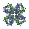
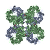
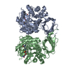
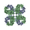
- Assembly
| Deposited unit | 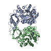
| ||||||||
|---|---|---|---|---|---|---|---|---|---|
| 1 | 
| ||||||||
| Unit cell |
| ||||||||
| Components on special symmetry positions |
| ||||||||
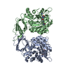
| Details | homo-octomer. Remainder of biological assembly generated by rotation about the 4-fold axis at 1/2,0,z (59.5, 0, 37.55) |
-Components
| #1: Protein | Mass: 41166.773 Da / Num. of mol.: 2 / Mutation: E77Q Source method: isolated from a genetically manipulated source Source: (gene. exp.) References: UniProt: P39377, Hydrolases; Acting on peptide bonds (peptidases); Omega peptidases #2: Chemical | ChemComp-ZN /   Mass: 65.409 Da / Num. of mol.: 4 / Source method: obtained synthetically / Formula: Zn Mass: 65.409 Da / Num. of mol.: 4 / Source method: obtained synthetically / Formula: Zn#3: Water | ChemComp-HOH / |  Mass: 18.015 Da / Num. of mol.: 422 / Source method: isolated from a natural source / Formula: H2O Mass: 18.015 Da / Num. of mol.: 422 / Source method: isolated from a natural source / Formula: H2O |
|---|
-Experimental details
-Experiment
| Experiment | Method: X-RAY DIFFRACTION / Number of used crystals: 1 |
|---|
- Sample preparation
| Crystal | Density Matthews: 2.9 Å3/Da / Density % sol: 57 % |
|---|---|
| Crystal grow | Temperature: 290 K / Method: vapor diffusion, hanging drop / pH: 5 Details: PEG-8000, MgCl2, homopipes, pH 5.0, VAPOR DIFFUSION, HANGING DROP, temperature 290K |
-Data collection
| Diffraction | Mean temperature: 277 K |
|---|---|
| Diffraction source | Source: ROTATING ANODE / Type: RIGAKU RU200 / Wavelength: 1.5418 Å |
| Detector | Type: SIEMENS HI-STAR / Detector: AREA DETECTOR / Date: Aug 20, 2003 / Details: supper mirrors |
| Radiation | Monochromator: Ni filter / Protocol: SINGLE WAVELENGTH / Monochromatic (M) / Laue (L): M / Scattering type: x-ray |
| Radiation wavelength | Wavelength: 1.5418 Å / Relative weight: 1 |
| Reflection | Resolution: 1.95→30 Å / Num. all: 68348 / Num. obs: 68348 / % possible obs: 93.4 % / Observed criterion σ(F): 0 / Observed criterion σ(I): 0 / Redundancy: 3.7 % / Rsym value: 0.07 / Net I/σ(I): 8.2 |
| Reflection shell | Resolution: 1.95→2.04 Å / Redundancy: 1.7 % / Mean I/σ(I) obs: 1.7 / Num. unique all: 6900 / Rsym value: 0.28 / % possible all: 75.7 |
- Processing
| Software |
| |||||||||||||||||||||||||
|---|---|---|---|---|---|---|---|---|---|---|---|---|---|---|---|---|---|---|---|---|---|---|---|---|---|---|
| Refinement | Method to determine structure: FOURIER SYNTHESIS Starting model: PDB entry 1onw Resolution: 1.95→30 Å / Cross valid method: THROUGHOUT / σ(F): 0 / Stereochemistry target values: Engh & Huber
| |||||||||||||||||||||||||
| Displacement parameters | Biso mean: 29.604 Å2 | |||||||||||||||||||||||||
| Refinement step | Cycle: LAST / Resolution: 1.95→30 Å
|
