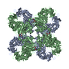
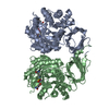
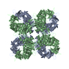
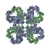
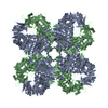
The biological assembly is an octomer. It is generated by expanding the crystallographically independent unit around the 4-fold crystallographic axis. The three rotational & translational matrices used to generate the octamer are as follows: (I) TRANSLATION VECTOR IN fractions of cell edge 0.500 0.500 0.000 ROTATION MATRIX 0.000 -1.000 0.000 1.000 0.000 0.000 0.000 0.000 1.000 (II) TRANSLATION VECTOR IN fractions of cell edge 0.000 1.000 0.000 ROTATION MATRIX -1.000 0.000 0.000 0.000 -1.000 0.000 0.000 0.000 1.000 TRANSLATION VECTOR IN fractions of cell edge -0.500 0.500 0.000 ROTATION MATRIX 0.000 1.000 0.000 -1.000 0.000 0.000 0.000 0.000 1.000 (III)TRANSLATION VECTOR IN fractions of cell edge -0.500 0.500 0.000 ROTATION MATRIX 0.000 1.000 0.000 -1.000 0.000 0.000 0.000 0.000 1.000
-
Components
-
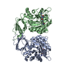
Protein , 1 types, 2 molecules AB
#1: Protein
Isoaspartyldipeptidase
Mass: 41167.758 Da / Num. of mol.: 2 Source method: isolated from a genetically manipulated source Source: (gene. exp.) Escherichia coli (E. coli) / Gene: IADA OR B4328 / Production host: Escherichia coli (E. coli) / Strain (production host): BL21(DE3)star References: UniProt: P39377, Hydrolases; Acting on peptide bonds (peptidases); Omega peptidases
In the structure databanks used in Yorodumi, some data are registered as the other names, "COVID-19 virus" and "2019-nCoV". Here are the details of the virus and the list of structure data.
Jan 31, 2019. EMDB accession codes are about to change! (news from PDBe EMDB page)
EMDB accession codes are about to change! (news from PDBe EMDB page)
The allocation of 4 digits for EMDB accession codes will soon come to an end. Whilst these codes will remain in use, new EMDB accession codes will include an additional digit and will expand incrementally as the available range of codes is exhausted. The current 4-digit format prefixed with “EMD-” (i.e. EMD-XXXX) will advance to a 5-digit format (i.e. EMD-XXXXX), and so on. It is currently estimated that the 4-digit codes will be depleted around Spring 2019, at which point the 5-digit format will come into force.
The EM Navigator/Yorodumi systems omit the EMD- prefix.
Related info.:Q: What is EMD? / ID/Accession-code notation in Yorodumi/EM Navigator
Yorodumi is a browser for structure data from EMDB, PDB, SASBDB, etc.
This page is also the successor to EM Navigator detail page, and also detail information page/front-end page for Omokage search.
The word "yorodu" (or yorozu) is an old Japanese word meaning "ten thousand". "mi" (miru) is to see.
Related info.:EMDB / PDB / SASBDB / Comparison of 3 databanks / Yorodumi Search / Aug 31, 2016. New EM Navigator & Yorodumi / Yorodumi Papers / Jmol/JSmol / Function and homology information / Changes in new EM Navigator and Yorodumi
 Movie
Movie Controller


 Open data
 Basic information
 Components
 Keywords
 Function and homology information
 X-RAY DIFFRACTION /
  Authors
 Citation
 Structure visualization
 Downloads & links
 Other downloads


 PDBj
PDBj

 Assembly
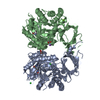


 Mass: 65.409 Da / Num. of mol.: 4 / Source method: obtained synthetically / Formula: Zn
 Mass: 35.453 Da / Num. of mol.: 3 / Source method: obtained synthetically / Formula: Cl
 Mass: 62.068 Da / Num. of mol.: 3 / Source method: obtained synthetically / Formula: C2H6O2
 Mass: 24.305 Da / Num. of mol.: 1 / Source method: obtained synthetically / Formula: Mg
 Mass: 22.990 Da / Num. of mol.: 1 / Source method: obtained synthetically / Formula: Na
 Sample preparation
 Processing