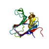
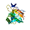
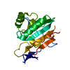
- PDB-2al7: Structure Of Human ADP-Ribosylation Factor-Like 10C -
+
Open data
ID or keywords:
Loading...
-
Basic information
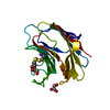
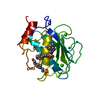
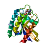
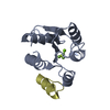
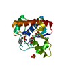
Entry
Database: PDB / ID: 2al7
Title
Structure Of Human ADP-Ribosylation Factor-Like 10C
Components
ADP-ribosylation factor-like 10C
Keywords
TRANSPORT PROTEIN / GDP-BINDING / MEMBRANE TRAFFICKING / STRUCTURAL GENOMICS / STRUCTURAL GENOMICS CONSORTIUM / SGC
Function / homology
Function and homology information
antigen processing and presentation of polysaccharide antigen via MHC class II / antigen processing and presentation following phagocytosis / viral exocytosis / calcium ion regulated lysosome exocytosis / endosome to lysosome transport of low-density lipoprotein particle / early endosome to Golgi transport / phagosome-lysosome fusion / protein localization to early endosome / cytolytic granule membrane / late endosome to lysosome transport ...antigen processing and presentation of polysaccharide antigen via MHC class II / antigen processing and presentation following phagocytosis / viral exocytosis / calcium ion regulated lysosome exocytosis / endosome to lysosome transport of low-density lipoprotein particle / early endosome to Golgi transport / phagosome-lysosome fusion / protein localization to early endosome / cytolytic granule membrane / late endosome to lysosome transport / autophagosome-lysosome fusion / lysosome localization / anterograde axonal transport / plasma membrane repair / natural killer cell mediated cytotoxicity / spindle midzone / alpha-tubulin binding / beta-tubulin binding / axon cytoplasm / small monomeric GTPase / chromosome segregation / GDP binding / late endosome membrane / protein transport / G protein activity / early endosome membrane / midbody / lysosome / cell division / lysosomal membrane / GTPase activity / synapse / GTP binding / extracellular exosome / membrane / cytosol / cytoplasm Similarity search - Function
ADP-ribosylation factor-like protein 8A/8B / Small GTPase Arf domain profile. / Sar1p-like members of the Ras-family of small GTPases / Small GTPase superfamily, ARF/SAR type / ADP-ribosylation factor family / ARF-like small GTPases; ARF, ADP-ribosylation factor / Ras subfamily of RAS small GTPases / Rab subfamily of small GTPases / Small GTP-binding protein domain / P-loop containing nucleotide triphosphate hydrolases ...ADP-ribosylation factor-like protein 8A/8B / Small GTPase Arf domain profile. / Sar1p-like members of the Ras-family of small GTPases / Small GTPase superfamily, ARF/SAR type / ADP-ribosylation factor family / ARF-like small GTPases; ARF, ADP-ribosylation factor / Ras subfamily of RAS small GTPases / Rab subfamily of small GTPases / Small GTP-binding protein domain / P-loop containing nucleotide triphosphate hydrolases / Rossmann fold / P-loop containing nucleoside triphosphate hydrolase / 3-Layer(aba) Sandwich / Alpha Beta Similarity search - Domain/homology
In the structure databanks used in Yorodumi, some data are registered as the other names, "COVID-19 virus" and "2019-nCoV". Here are the details of the virus and the list of structure data.
Jan 31, 2019. EMDB accession codes are about to change! (news from PDBe EMDB page)
EMDB accession codes are about to change! (news from PDBe EMDB page)
The allocation of 4 digits for EMDB accession codes will soon come to an end. Whilst these codes will remain in use, new EMDB accession codes will include an additional digit and will expand incrementally as the available range of codes is exhausted. The current 4-digit format prefixed with “EMD-” (i.e. EMD-XXXX) will advance to a 5-digit format (i.e. EMD-XXXXX), and so on. It is currently estimated that the 4-digit codes will be depleted around Spring 2019, at which point the 5-digit format will come into force.
The EM Navigator/Yorodumi systems omit the EMD- prefix.
Related info.:Q: What is EMD? / ID/Accession-code notation in Yorodumi/EM Navigator
Yorodumi is a browser for structure data from EMDB, PDB, SASBDB, etc.
This page is also the successor to EM Navigator detail page, and also detail information page/front-end page for Omokage search.
The word "yorodu" (or yorozu) is an old Japanese word meaning "ten thousand". "mi" (miru) is to see.
Related info.:EMDB / PDB / SASBDB / Comparison of 3 databanks / Yorodumi Search / Aug 31, 2016. New EM Navigator & Yorodumi / Yorodumi Papers / Jmol/JSmol / Function and homology information / Changes in new EM Navigator and Yorodumi
 Movie
Movie Controller


 Open data
 Basic information
 Components
 Keywords
 Function and homology information
 Homo sapiens (human)
 X-RAY DIFFRACTION /
  Authors
 Citation
 Structure visualization
 Downloads & links
 Other downloads
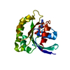


 PDBj
PDBj


 Assembly


 Mass: 24.305 Da / Num. of mol.: 1 / Source method: obtained synthetically / Formula: Mg
 Type: RNA linking / Mass: 443.201 Da / Num. of mol.: 1 / Source method: obtained synthetically / Formula: C10H15N5O11P2 / Comment: GDP, energy-carrying molecule*YM
 Mass: 18.015 Da / Num. of mol.: 142 / Source method: isolated from a natural source / Formula: H2O
 Sample preparation
 Processing