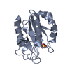
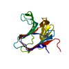
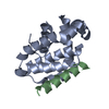
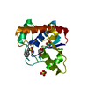
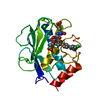
Entry Database : PDB / ID : 4rsgTitle Neutron crystal structure of Ras bound to the GTP analogue GppNHp GTPase HRas Keywords / / / Function / homology Function Domain/homology Component
/ / / / / / / / / / / / / / / / / / / / / / / / / / / / / / / / / / / / / / / / / / / / / / / / / / / / / / / / / / / / / / / / / / / / / / / / / / / / / / / / / / / / / / / / / / / / / / / / / / / / / / / / / / / / / / / / / / / / / / / / / / / / / / / / / Biological species Homo sapiens (human)Method / / / Resolution : 1.907 Å Authors Knihtila, R.R. / Holzapfel, G. / Weiss, K.L. / Meilleur, F. / Mattos, C. Journal : J.Biol.Chem. / Year : 2015Title : Neutron Crystal Structure of RAS GTPase Puts in Question the Protonation State of the GTP gamma-Phosphate.Authors : Knihtila, R. / Holzapfel, G. / Weiss, K. / Meilleur, F. / Mattos, C. History Deposition Nov 7, 2014 Deposition site / Processing site Revision 1.0 Nov 4, 2015 Provider / Type Revision 1.1 Nov 18, 2015 Group Revision 1.2 Jan 13, 2016 Group Revision 1.3 Apr 25, 2018 Group / Category Item _diffrn_source.pdbx_synchrotron_beamline / _diffrn_source.pdbx_synchrotron_site ... _diffrn_source.pdbx_synchrotron_beamline / _diffrn_source.pdbx_synchrotron_site / _diffrn_source.source / _diffrn_source.type Revision 1.4 Jun 30, 2021 Group / Derived calculationsCategory diffrn_detector / diffrn_radiation ... diffrn_detector / diffrn_radiation / pdbx_struct_conn_angle / struct_conn / struct_site Item _diffrn_detector.detector / _diffrn_detector.type ... _diffrn_detector.detector / _diffrn_detector.type / _diffrn_radiation.pdbx_monochromatic_or_laue_m_l / _pdbx_struct_conn_angle.ptnr1_auth_comp_id / _pdbx_struct_conn_angle.ptnr1_auth_seq_id / _pdbx_struct_conn_angle.ptnr1_label_asym_id / _pdbx_struct_conn_angle.ptnr1_label_atom_id / _pdbx_struct_conn_angle.ptnr1_label_comp_id / _pdbx_struct_conn_angle.ptnr1_label_seq_id / _pdbx_struct_conn_angle.ptnr3_auth_comp_id / _pdbx_struct_conn_angle.ptnr3_auth_seq_id / _pdbx_struct_conn_angle.ptnr3_label_asym_id / _pdbx_struct_conn_angle.ptnr3_label_atom_id / _pdbx_struct_conn_angle.ptnr3_label_comp_id / _pdbx_struct_conn_angle.ptnr3_label_seq_id / _pdbx_struct_conn_angle.value / _struct_conn.pdbx_dist_value / _struct_conn.ptnr1_auth_comp_id / _struct_conn.ptnr1_auth_seq_id / _struct_conn.ptnr1_label_asym_id / _struct_conn.ptnr1_label_atom_id / _struct_conn.ptnr1_label_comp_id / _struct_conn.ptnr1_label_seq_id / _struct_conn.ptnr2_auth_comp_id / _struct_conn.ptnr2_auth_seq_id / _struct_conn.ptnr2_label_asym_id / _struct_conn.ptnr2_label_atom_id / _struct_conn.ptnr2_label_comp_id / _struct_site.pdbx_auth_asym_id / _struct_site.pdbx_auth_comp_id / _struct_site.pdbx_auth_seq_id Revision 1.5 Feb 28, 2024 Group / Database references / Category / chem_comp_bond / database_2Item / _database_2.pdbx_database_accession
Show all Show less
 Movie
Movie Controller

 Yorodumi
 Open data
 Basic information
 Components
 Keywords
 Function and homology information
 Homo sapiens (human)
 MOLECULAR REPLACEMENT / Resolution: 1.907 Å
  Authors
 Citation
 Structure visualization
 Downloads & links
 Other downloads
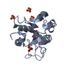


 PDBj
PDBj


 Assembly
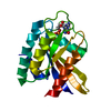


 Mass: 522.196 Da / Num. of mol.: 1 / Source method: obtained synthetically / Formula: C10H17N6O13P3
 Mass: 24.305 Da / Num. of mol.: 1 / Source method: obtained synthetically / Formula: Mg
 Mass: 18.015 Da / Num. of mol.: 75 / Source method: isolated from a natural source / Formula: D2O
 Sample preparation
 / Beamline: CG4D / Wavelength: 3.3-4.5
 Processing