 Movie
Movie Controller

[English] 日本語
 Yorodumi
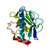
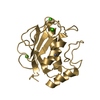
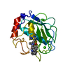
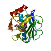
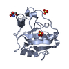
- PDB-5cxa: Crystal structure of the catalytic domain of Human MMP12 in compl... -
+ Open data

- Basic information
| Entry | Database: PDB / ID: 5cxa | ||||||
|---|---|---|---|---|---|---|---|
| Title | Crystal structure of the catalytic domain of Human MMP12 in complex with a carboxylate inhibitor related to RXP470 | ||||||
 Components Components | Macrophage metalloelastase | ||||||
 Keywords Keywords | HYDROLASE / metzincin / RXP470 / carboxylate based inhibitor / carboxylate peptidic inhibitor | ||||||
| Function / homology |  Function and homology information Function and homology informationmacrophage elastase / negative regulation of endothelial cell-matrix adhesion / bronchiole development / positive regulation of epithelial cell proliferation involved in wound healing / elastin catabolic process / regulation of defense response to virus by host / positive regulation of type I interferon-mediated signaling pathway / wound healing, spreading of epidermal cells / negative regulation of type I interferon-mediated signaling pathway / lung alveolus development ...macrophage elastase / negative regulation of endothelial cell-matrix adhesion / bronchiole development / positive regulation of epithelial cell proliferation involved in wound healing / elastin catabolic process / regulation of defense response to virus by host / positive regulation of type I interferon-mediated signaling pathway / wound healing, spreading of epidermal cells / negative regulation of type I interferon-mediated signaling pathway / lung alveolus development / response to amyloid-beta / Collagen degradation / collagen catabolic process / positive regulation of interferon-alpha production / extracellular matrix disassembly / core promoter sequence-specific DNA binding / collagen binding / Degradation of the extracellular matrix / extracellular matrix organization / metalloendopeptidase activity / cellular response to virus / protein import into nucleus / extracellular matrix / endopeptidase activity / sequence-specific DNA binding / serine-type endopeptidase activity / calcium ion binding / negative regulation of transcription by RNA polymerase II / positive regulation of transcription by RNA polymerase II / proteolysis / : / extracellular region / zinc ion binding / nucleus / cytoplasm Similarity search - Function | ||||||
| Biological species |  Homo sapiens (human) Homo sapiens (human) | ||||||
| Method |  X-RAY DIFFRACTION / SYNCHROTRON / MOLECULAR REPLACEMENT / Resolution: 1.3 Å X-RAY DIFFRACTION / SYNCHROTRON / MOLECULAR REPLACEMENT / Resolution: 1.3 Å | ||||||
 Authors Authors | Rouanet-Mehouas, C. / Devel, L. / Dive, V. / Stura, E.A. | ||||||
 Citation Citation | Journal: J. Med. Chem. / Year: 2017 Title: Zinc-Metalloproteinase Inhibitors: Evaluation of the Complex Role Played by the Zinc-Binding Group on Potency and Selectivity. Authors: Rouanet-Mehouas, C. / Czarny, B. / Beau, F. / Cassar-Lajeunesse, E. / Stura, E.A. / Dive, V. / Devel, L. | ||||||
| History |
|
- Structure visualization
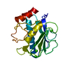
| Structure viewer | Molecule: MolmilJmol/JSmol |
|---|
- Downloads & links
-Download
| PDBx/mmCIF format | 5cxa.cif.gz | 97.6 KB | Display | PDBx/mmCIF format |
|---|---|---|---|---|
| PDB format | pdb5cxa.ent.gz | 73.5 KB | Display | PDB format |
| PDBx/mmJSON format | 5cxa.json.gz | Tree view | PDBx/mmJSON format | |
| Others |  Other downloads Other downloads |
-Validation report
| Arichive directory | https://data.pdbj.org/pub/pdb/validation_reports/cx/5cxaftp://data.pdbj.org/pub/pdb/validation_reports/cx/5cxa | HTTPS FTP |
|---|
-Related structure data
| Related structure data |  5czmC  5d2bC  5d3cC  4gqlS S: Starting model for refinement C: citing same article ( |
|---|---|
| Similar structure data |


-Links
 PDBj
PDBj


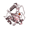
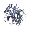
- Assembly
| Deposited unit | 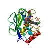
| ||||||||||||
|---|---|---|---|---|---|---|---|---|---|---|---|---|---|
| 1 |
| ||||||||||||
| Unit cell |
| ||||||||||||
| Components on special symmetry positions |
|
-Components
| #1: Protein | Mass: 17557.568 Da / Num. of mol.: 1 / Fragment: UNP residues 106-263 / Mutation: F171D, K241A Source method: isolated from a genetically manipulated source Details: MMP12 F171D K241A mutant for calorimetric studies / Source: (gene. exp.) Homo sapiens (human) / Gene: MMP12, HME / Plasmid: PET24A / Production host:  | ||||||
|---|---|---|---|---|---|---|---|
| #2: Chemical |   Mass: 65.409 Da / Num. of mol.: 2 / Source method: obtained synthetically / Formula: Zn Mass: 65.409 Da / Num. of mol.: 2 / Source method: obtained synthetically / Formula: Zn#3: Chemical |   Mass: 40.078 Da / Num. of mol.: 3 / Source method: obtained synthetically / Formula: Ca Mass: 40.078 Da / Num. of mol.: 3 / Source method: obtained synthetically / Formula: Ca#4: Chemical | ChemComp-55L / ( | 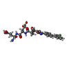  Mass: 643.041 Da / Num. of mol.: 1 / Fragment: UNP residues 107-263 / Mutation: F171D, K241A Mass: 643.041 Da / Num. of mol.: 1 / Fragment: UNP residues 107-263 / Mutation: F171D, K241ASource method: isolated from a genetically manipulated source Formula: C30H31ClN4O10 / Details: MMP12 F171D K241A mutant for calorimetric studies / Source: (gene. exp.) Homo sapiens (human) / Gene: MMP12, HME / Plasmid: PET24A / Production host: #5: Water | ChemComp-HOH / |  Mass: 18.015 Da / Num. of mol.: 285 / Source method: isolated from a natural source / Formula: H2O Mass: 18.015 Da / Num. of mol.: 285 / Source method: isolated from a natural source / Formula: H2O |
-Experimental details
-Experiment
| Experiment | Method: X-RAY DIFFRACTION / Number of used crystals: 1 |
|---|
- Sample preparation
| Crystal | Density Matthews: 2.29 Å3/Da / Density % sol: 46.18 % / Description: prismatic |
|---|---|
| Crystal grow | Temperature: 293 K / Method: vapor diffusion, sitting drop / pH: 10 Details: Protein: hMMP12 F171D K241A at 608 micro-M + 10 milli-M AHA + 0.552 milli-M inhibitor. precipitant: 26% PEG4000, 3% dioxane, 0.114 M TRIS pH 10. cryoprotectant: 10 % diethylene glycol + 5 % ...Details: Protein: hMMP12 F171D K241A at 608 micro-M + 10 milli-M AHA + 0.552 milli-M inhibitor. precipitant: 26% PEG4000, 3% dioxane, 0.114 M TRIS pH 10. cryoprotectant: 10 % diethylene glycol + 5 % glycerol + 10 % 2,3-butanediol + 5 % 1,4-dioxane, 25% PEG 6K, 0.1 M TRIS-HCl, pH 8.0 PH range: 8-10 / Temp details: cooled incubator |
-Data collection
| Diffraction | Mean temperature: 100 K / Ambient temp details: N2 stream |
|---|---|
| Diffraction source | Source: SYNCHROTRON / Site: ESRF  / Beamline: ID23-2 / Wavelength: 0.8729 Å / Beamline: ID23-2 / Wavelength: 0.8729 Å |
| Detector | Type: DECTRIS PILATUS 6M / Detector: PIXEL / Date: Sep 7, 2014 / Details: bent cylindrical mirror |
| Radiation | Monochromator: horizontally diffracting Si (111) monochromator and Pt coated mirrors in Kirkpatrick-Baez geometry for focusing Protocol: SINGLE WAVELENGTH / Monochromatic (M) / Laue (L): M / Scattering type: x-ray |
| Radiation wavelength | Wavelength: 0.8729 Å / Relative weight: 1 |
| Reflection | Resolution: 1.3→46.88 Å / Num. all: 40627 / Num. obs: 40584 / % possible obs: 99.9 % / Observed criterion σ(F): 0 / Observed criterion σ(I): -3 / Redundancy: 9.05 % / Rmerge(I) obs: 0.165 / Rsym value: 0.155 / Net I/σ(I): 9.23 |
| Reflection shell | Resolution: 1.3→1.38 Å / Redundancy: 8.67 % / Rmerge(I) obs: 1.39 / Mean I/σ(I) obs: 1.24 / % possible all: 99.3 |
- Processing
| Software |
| |||||||||||||||||||||||||||||||||||||||||||||||||||||||||||||||||||||||||||||||||||||||||||||||||||||||||
|---|---|---|---|---|---|---|---|---|---|---|---|---|---|---|---|---|---|---|---|---|---|---|---|---|---|---|---|---|---|---|---|---|---|---|---|---|---|---|---|---|---|---|---|---|---|---|---|---|---|---|---|---|---|---|---|---|---|---|---|---|---|---|---|---|---|---|---|---|---|---|---|---|---|---|---|---|---|---|---|---|---|---|---|---|---|---|---|---|---|---|---|---|---|---|---|---|---|---|---|---|---|---|---|---|---|---|
| Refinement | Method to determine structure: MOLECULAR REPLACEMENT Starting model: 4GQL Resolution: 1.3→36.62 Å / SU ML: 0.14 / Cross valid method: THROUGHOUT / σ(F): 1.35 / Phase error: 17.84 / Stereochemistry target values: ML
| |||||||||||||||||||||||||||||||||||||||||||||||||||||||||||||||||||||||||||||||||||||||||||||||||||||||||
| Solvent computation | Shrinkage radii: 0.9 Å / VDW probe radii: 1.11 Å / Solvent model: FLAT BULK SOLVENT MODEL | |||||||||||||||||||||||||||||||||||||||||||||||||||||||||||||||||||||||||||||||||||||||||||||||||||||||||
| Refinement step | Cycle: LAST / Resolution: 1.3→36.62 Å
| |||||||||||||||||||||||||||||||||||||||||||||||||||||||||||||||||||||||||||||||||||||||||||||||||||||||||
| Refine LS restraints |
| |||||||||||||||||||||||||||||||||||||||||||||||||||||||||||||||||||||||||||||||||||||||||||||||||||||||||
| LS refinement shell |
|
