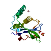
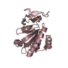
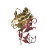
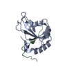
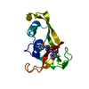
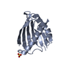
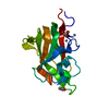
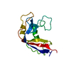
#1: Journal: J.Mol.Biol. / Year: 2004 Title: Structure of human Ki67 FHA domain and its binding to a phosphoprotein fragment from hNIFK reveal unique recognition sites and new views to the structural basis of FHA domain functions. Authors: Li, H. / Byeon, I.-J.L. / Ju, Y. / Tsai, M.-D.
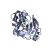
structures with the least restraint violations,structures with the lowest energy
Representative
Model #1
closest to the average
-
Components
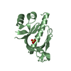
#1: Protein
AntigenKI-67
Mass: 13826.766 Da / Num. of mol.: 1 / Fragment: FHA domain Source method: isolated from a genetically manipulated source Source: (gene. exp.) Homo sapiens (human) / Genus: Escherichia / Gene: MKI67 / Plasmid: PEG / Species (production host): Escherichia coli / Production host: Escherichia coli BL21(DE3) (bacteria) / Strain (production host): BL21(DE3) / References: UniProt: P46013
#2: Protein/peptide
MKI67FHAdomaininteractingnucleolarphosphoprotein / Nucleolar protein interacting with the FHA domain of pKI-67 / hNIFK / Nucleolar phosphoprotein Nopp34
Mass: 5197.442 Da / Num. of mol.: 1 / Fragment: residues 226-269 Source method: isolated from a genetically manipulated source Source: (gene. exp.) Homo sapiens (human) / Genus: Escherichia / Gene: MKI67IP / Plasmid: pEG / Species (production host): Escherichia coli / Production host: Escherichia coli BL21(DE3) (bacteria) / Strain (production host): BL21(DE3) / References: UniProt: Q9BYG3
Has protein modification
Y
-
Experimental details
-
Experiment
Experiment
Method: SOLUTION NMR
NMR experiment
Conditions-ID
Experiment-ID
Solution-ID
Type
1
1
1
3D 13C-separated NOESY
1
2
1
3D 15N-separated NOESY
1
3
3
3D 13C-separated NOESY
1
4
3
3D 15N-separated NOESY
1
5
1
3D 12C/14N-filtered 13C separated NOESY
1
6
3
3D 12C/14N-filtered 13C separated NOESY
NMR details
Text: The structure was determined using triple-resonance NMR spectroscopy
-
Sample preparation
Details
Solution-ID
Contents
Solvent system
1
0.9 mM Ki67 FHA U-15N,13C; 1 mM hNIFK(226-269)3P unlabeled 5 mM HEPES, 5 mM DTT, 1 mM EDTA, 150 mM NaCl, pH 7.4
93% H2O/7% D2O
2
0.9 mM Ki67 FHA U-15N,13C; 1 mM hNIFK(226-269)3P unlabeled 5 mM HEPES, 5 mM DTT, 1 mM EDTA, 150 mM NaCl, pH 7.4
100% D2O
3
1 mM Ki67 FHA unlabled; 0.9 mM hNIFK(226-269)3P U-15N,13C 5 mM HEPES, 5 mM DTT, 1 mM EDTA, 150 mM NaCl, pH 7.4
93% H2O/7% D2O
4
1 mM Ki67 FHA unlabled; 0.9 mM hNIFK(226-269)3P U-15N,13C 5 mM HEPES, 5 mM DTT, 1 mM EDTA, 150 mM NaCl, pH 7.4
100% D2O
Sample conditions
Ionic strength: 150 mM NaCl / pH: 7.4 / Pressure: ambient / Temperature: 293 K
-
NMR measurement
NMR spectrometer
Type
Manufacturer
Model
Field strength (MHz)
Spectrometer-ID
Bruker AVANCE
Bruker
AVANCE
800
1
Bruker DMX
Bruker
DMX
750
2
Bruker AVANCE
Bruker
AVANCE
600
3
Bruker DMX
Bruker
DMX
500
4
-
Processing
NMR software
Name
Version
Developer
Classification
X-PLOR
NIHversion
Brunger
structuresolution
X-PLOR
NIHversion
Brunger
refinement
Refinement
Method: simulated annealing / Software ordinal: 1 Details: the structures are based on a total of 3476 constraints, 3141 are distance, 215 dihedral angle, and 120 N-H residual dipolar coupling constraints
NMR representative
Selection criteria: closest to the average
NMR ensemble
Conformer selection criteria: structures with the least restraint violations,structures with the lowest energy Conformers calculated total number: 512 / Conformers submitted total number: 100
+
About Yorodumi
-
News
-
Feb 9, 2022. New format data for meta-information of EMDB entries
New format data for meta-information of EMDB entries
Version 3 of the EMDB header file is now the official format.
The previous official version 1.9 will be removed from the archive.
In the structure databanks used in Yorodumi, some data are registered as the other names, "COVID-19 virus" and "2019-nCoV". Here are the details of the virus and the list of structure data.
Jan 31, 2019. EMDB accession codes are about to change! (news from PDBe EMDB page)
EMDB accession codes are about to change! (news from PDBe EMDB page)
The allocation of 4 digits for EMDB accession codes will soon come to an end. Whilst these codes will remain in use, new EMDB accession codes will include an additional digit and will expand incrementally as the available range of codes is exhausted. The current 4-digit format prefixed with “EMD-” (i.e. EMD-XXXX) will advance to a 5-digit format (i.e. EMD-XXXXX), and so on. It is currently estimated that the 4-digit codes will be depleted around Spring 2019, at which point the 5-digit format will come into force.
The EM Navigator/Yorodumi systems omit the EMD- prefix.
Related info.:Q: What is EMD? / ID/Accession-code notation in Yorodumi/EM Navigator
Yorodumi is a browser for structure data from EMDB, PDB, SASBDB, etc.
This page is also the successor to EM Navigator detail page, and also detail information page/front-end page for Omokage search.
The word "yorodu" (or yorozu) is an old Japanese word meaning "ten thousand". "mi" (miru) is to see.
Related info.:EMDB / PDB / SASBDB / Comparison of 3 databanks / Yorodumi Search / Aug 31, 2016. New EM Navigator & Yorodumi / Yorodumi Papers / Jmol/JSmol / Function and homology information / Changes in new EM Navigator and Yorodumi
 Movie
Movie Controller


 Open data
 Basic information
 Components
 Keywords
 Function and homology information
 Homo sapiens (human)
 Authors
 Citation
 Structure visualization
 Downloads & links
 Other downloads
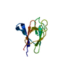
 PDBj
PDBj

 Assembly

 Sample preparation
 Processing
 X-PLOR