SOLUTION NMR / The structure calculations were made with the program CYANA 2.0 using the automated NOE assignment, structure calculation algorithm. The structure refinement of the 20 final CYANA structures was made with AMBER 8.0 program using the generalized Born continuum solvent model.
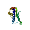
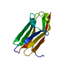
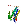
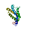
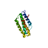
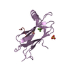
FilaminA / Alpha-filamin / Filamin 1 / Endothelial actin-binding protein / Actin-binding protein 280 / ABP-280 ...Alpha-filamin / Filamin 1 / Endothelial actin-binding protein / Actin-binding protein 280 / ABP-280 / Nonmuscle filamin
Mass: 10075.146 Da / Num. of mol.: 1 / Fragment: Filamin A domain 17 Source method: isolated from a genetically manipulated source Source: (gene. exp.) Homo sapiens (human) / Production host: Escherichia coli (E. coli) / References: UniProt: P21333
-
Experimental details
-
Experiment
Experiment
Method: SOLUTION NMR
NMR experiment
Conditions-ID
Experiment-ID
Solution-ID
Type
1
1
1
3D 13C-separated NOESY
1
2
1
3D 15N-separated NOESY
NMR details
Text: The numbering of residues in constraint and chemical shift files corresponds to the numbering of NMR construct (Residues 1-98).
Method: The structure calculations were made with the program CYANA 2.0 using the automated NOE assignment, structure calculation algorithm. The structure refinement of the 20 final CYANA structures ...Method: The structure calculations were made with the program CYANA 2.0 using the automated NOE assignment, structure calculation algorithm. The structure refinement of the 20 final CYANA structures was made with AMBER 8.0 program using the generalized Born continuum solvent model. Software ordinal: 1
NMR representative
Selection criteria: closest to the average
NMR ensemble
Conformer selection criteria: structures with favorable non-bond energy Conformers calculated total number: 300 / Conformers submitted total number: 20
+
About Yorodumi
-
News
-
Feb 9, 2022. New format data for meta-information of EMDB entries
New format data for meta-information of EMDB entries
Version 3 of the EMDB header file is now the official format.
The previous official version 1.9 will be removed from the archive.
In the structure databanks used in Yorodumi, some data are registered as the other names, "COVID-19 virus" and "2019-nCoV". Here are the details of the virus and the list of structure data.
Jan 31, 2019. EMDB accession codes are about to change! (news from PDBe EMDB page)
EMDB accession codes are about to change! (news from PDBe EMDB page)
The allocation of 4 digits for EMDB accession codes will soon come to an end. Whilst these codes will remain in use, new EMDB accession codes will include an additional digit and will expand incrementally as the available range of codes is exhausted. The current 4-digit format prefixed with “EMD-” (i.e. EMD-XXXX) will advance to a 5-digit format (i.e. EMD-XXXXX), and so on. It is currently estimated that the 4-digit codes will be depleted around Spring 2019, at which point the 5-digit format will come into force.
The EM Navigator/Yorodumi systems omit the EMD- prefix.
Related info.:Q: What is EMD? / ID/Accession-code notation in Yorodumi/EM Navigator
Yorodumi is a browser for structure data from EMDB, PDB, SASBDB, etc.
This page is also the successor to EM Navigator detail page, and also detail information page/front-end page for Omokage search.
The word "yorodu" (or yorozu) is an old Japanese word meaning "ten thousand". "mi" (miru) is to see.
Related info.:EMDB / PDB / SASBDB / Comparison of 3 databanks / Yorodumi Search / Aug 31, 2016. New EM Navigator & Yorodumi / Yorodumi Papers / Jmol/JSmol / Function and homology information / Changes in new EM Navigator and Yorodumi
 Movie
Movie Controller


 Open data
 Basic information
 Components
 Keywords
 Function and homology information
 Homo sapiens (human)
 Authors
 Citation
 Structure visualization
 Downloads & links
 Other downloads


 PDBj
PDBj


 Assembly
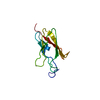

 Sample preparation
 Processing
 CYANA
CYANA