[English] 日本語
 Yorodumi
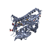
- PDB-2a8t: 2.1 Angstrom Crystal Structure of the Complex Between the Nuclear... -
+ Open data
- Basic information
| Entry | Database: PDB / ID: 2a8t | ||||||
|---|---|---|---|---|---|---|---|
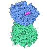
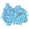
| Title | 2.1 Angstrom Crystal Structure of the Complex Between the Nuclear U8 snoRNA Decapping Nudix Hydrolase X29, Manganese and m7G-PPP-A | ||||||
 Components Components | U8 snoRNA-binding protein X29 | ||||||
 Keywords Keywords | TRANSLATION / HYDROLASE / modified nudix hydrolase fold | ||||||
| Function / homology |  Function and homology information Function and homology informationinosine diphosphate phosphatase / sno(s)RNA catabolic process / dIDP phosphatase activity / dITP catabolic process / IDP phosphatase activity / positive regulation of cell cycle process / RNA NAD+-cap (NAD+-forming) hydrolase activity / dITP diphosphatase activity / negative regulation of rRNA processing / phosphodiesterase decapping endonuclease activity ...inosine diphosphate phosphatase / sno(s)RNA catabolic process / dIDP phosphatase activity / dITP catabolic process / IDP phosphatase activity / positive regulation of cell cycle process / RNA NAD+-cap (NAD+-forming) hydrolase activity / dITP diphosphatase activity / negative regulation of rRNA processing / phosphodiesterase decapping endonuclease activity / 5'-(N7-methylguanosine 5'-triphospho)-[mRNA] hydrolase / NAD-cap decapping / 5'-(N(7)-methylguanosine 5'-triphospho)-[mRNA] hydrolase activity / metalloexopeptidase activity / cobalt ion binding / snoRNA binding / mRNA catabolic process / manganese ion binding / nucleotide binding / mRNA binding / nucleolus / magnesium ion binding / protein homodimerization activity / nucleoplasm / nucleus / cytoplasm Similarity search - Function | ||||||
| Biological species | |||||||
| Method |  X-RAY DIFFRACTION / X-RAY DIFFRACTION /  MOLECULAR REPLACEMENT / Resolution: 2.1 Å MOLECULAR REPLACEMENT / Resolution: 2.1 Å | ||||||
 Authors Authors | Scarsdale, J.N. / Peculis, B.A. / Wright, H.T. | ||||||
 Citation Citation |  Journal: Structure / Year: 2006 Journal: Structure / Year: 2006Title: Crystal structures of U8 snoRNA decapping nudix hydrolase, X29, and its metal and cap complexes Authors: Scarsdale, J.N. / Peculis, B.A. / Wright, H.T. #1:  Journal: Acta Crystallogr.,Sect.D / Year: 2004 Journal: Acta Crystallogr.,Sect.D / Year: 2004Title: Crystals of X29, a Xenopus Laevis U8 SnoRNA Binding Protein with Nuclear Decapping Activity Authors: Peculis, B.A. / Scarsdale, J.N. / Wright, H.T. #2:  Journal: Mol.Cell / Year: 2004 Journal: Mol.Cell / Year: 2004Title: Xenopus U8 SnoRNA Binding Protein is a Conserved Nuclear Decapping Enzyme Authors: Ghosh, T. / Peterson, B. / Tomasevic, N. / Peculis, B.A. | ||||||
| History |
|
- Structure visualization
| Structure viewer | Molecule:  Molmil Molmil Jmol/JSmol Jmol/JSmol |
|---|
- Downloads & links
- Download
| PDBx/mmCIF format |  2a8t.cif.gz 2a8t.cif.gz | 95.3 KB | Display |  PDBx/mmCIF format PDBx/mmCIF format |
|---|---|---|---|---|
| PDB format |  pdb2a8t.ent.gz pdb2a8t.ent.gz | 71.2 KB | Display |  PDB format PDB format |
| PDBx/mmJSON format |  2a8t.json.gz 2a8t.json.gz | Tree view |  PDBx/mmJSON format PDBx/mmJSON format | |
| Others |  Other downloads Other downloads |
-Validation report
| Summary document |  2a8t_validation.pdf.gz 2a8t_validation.pdf.gz | 1.7 MB | Display |  wwPDB validaton report wwPDB validaton report |
|---|---|---|---|---|
| Full document |  2a8t_full_validation.pdf.gz 2a8t_full_validation.pdf.gz | 1.7 MB | Display | |
| Data in XML |  2a8t_validation.xml.gz 2a8t_validation.xml.gz | 18.1 KB | Display | |
| Data in CIF |  2a8t_validation.cif.gz 2a8t_validation.cif.gz | 24.4 KB | Display | |
| Arichive directory |  https://data.pdbj.org/pub/pdb/validation_reports/a8/2a8t https://data.pdbj.org/pub/pdb/validation_reports/a8/2a8t ftp://data.pdbj.org/pub/pdb/validation_reports/a8/2a8t ftp://data.pdbj.org/pub/pdb/validation_reports/a8/2a8t | HTTPS FTP |
-Related structure data
| Related structure data | 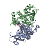 2a8pC 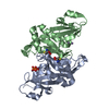 2a8qC 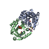 2a8rC 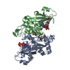 2a8sC 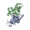 1u20S S: Starting model for refinement C: citing same article ( |
|---|---|
| Similar structure data |
- Links
Links
- Assembly
| Deposited unit | 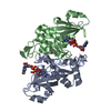
| ||||||||
|---|---|---|---|---|---|---|---|---|---|
| 1 |
| ||||||||
| Unit cell |
|
- Components
| #1: Protein | Mass: 24390.025 Da / Num. of mol.: 2 Source method: isolated from a genetically manipulated source Details: complexed with N-methyl-Guanosine-triphosphate-Guanosine Source: (gene. exp.)  References: UniProt: Q569R2, UniProt: Q6TEC1*PLUS, Hydrolases; Acting on acid anhydrides; In phosphorus-containing anhydrides #2: Chemical | ChemComp-MN / #3: Chemical | #4: Chemical | #5: Water | ChemComp-HOH / | |
|---|
-Experimental details
-Experiment
| Experiment | Method:  X-RAY DIFFRACTION / Number of used crystals: 1 X-RAY DIFFRACTION / Number of used crystals: 1 |
|---|
- Sample preparation
| Crystal | Density Matthews: 2.27 Å3/Da / Density % sol: 45.3 % |
|---|---|
| Crystal grow | Temperature: 293 K / pH: 7.68 Details: 4-5 mg/ml X29, 0.025M HEPES pH 7.68, 3/75% PEG 6000, VAPOR DIFFUSION, SITTING DROP, temperature 293K |
-Data collection
| Diffraction | Mean temperature: 95 K |
|---|---|
| Diffraction source | Source:  ROTATING ANODE / Type: RIGAKU MICROMAX-007 / Wavelength: 1.5418 ROTATING ANODE / Type: RIGAKU MICROMAX-007 / Wavelength: 1.5418 |
| Detector | Type: RIGAKU RAXIS IV / Detector: IMAGE PLATE / Date: Apr 3, 2005 / Details: OSMIC VARIMAX CONFOCAL OPTICS |
| Radiation | Monochromator: OSMIC VARIMAX CONFOCAL OPTICS / Protocol: SINGLE WAVELENGTH / Monochromatic (M) / Laue (L): M / Scattering type: x-ray |
| Radiation wavelength | Wavelength: 1.5418 Å / Relative weight: 1 |
| Reflection | Resolution: 2.1→46.17 Å / Num. obs: 27774 / % possible obs: 99.9 % / Redundancy: 6.92 % / Biso Wilson estimate: 57.6 Å2 / Rmerge(I) obs: 0.037 / Net I/σ(I): 19.8 |
| Reflection shell | Resolution: 2.1→2.18 Å / Redundancy: 6.88 % / Rmerge(I) obs: 0.38 / Mean I/σ(I) obs: 4.4 / % possible all: 100 |
- Processing
| Software |
| ||||||||||||||||||||||||||||||||||||||||||||||||||||||||||||||||||||||||||||||||||||||||||||||||||||||||||||||||||||||||||||||||||||||||||||||||||||||||||||||||||||||||||
|---|---|---|---|---|---|---|---|---|---|---|---|---|---|---|---|---|---|---|---|---|---|---|---|---|---|---|---|---|---|---|---|---|---|---|---|---|---|---|---|---|---|---|---|---|---|---|---|---|---|---|---|---|---|---|---|---|---|---|---|---|---|---|---|---|---|---|---|---|---|---|---|---|---|---|---|---|---|---|---|---|---|---|---|---|---|---|---|---|---|---|---|---|---|---|---|---|---|---|---|---|---|---|---|---|---|---|---|---|---|---|---|---|---|---|---|---|---|---|---|---|---|---|---|---|---|---|---|---|---|---|---|---|---|---|---|---|---|---|---|---|---|---|---|---|---|---|---|---|---|---|---|---|---|---|---|---|---|---|---|---|---|---|---|---|---|---|---|---|---|---|---|
| Refinement | Method to determine structure:  MOLECULAR REPLACEMENT MOLECULAR REPLACEMENTStarting model: PDB ENTRY 1U20 Resolution: 2.1→46.17 Å / Cor.coef. Fo:Fc: 0.96 / Cor.coef. Fo:Fc free: 0.938 / SU B: 12.983 / SU ML: 0.164 / TLS residual ADP flag: LIKELY RESIDUAL Isotropic thermal model: TLS refinement followed by restrained refinement of individual isotropic B factors Cross valid method: THROUGHOUT / σ(F): 0 / ESU R: 0.232 / ESU R Free: 0.203 / Stereochemistry target values: MAXIMUM LIKELIHOOD Details: SIMULATED ANNEALING VIA TORSION ANGLE DYNAMICS IN CNS V1.0 WAS FOLLOWED BY MAXIMUM LIKELIHOOD REFINEMENT IN REFMAC5
| ||||||||||||||||||||||||||||||||||||||||||||||||||||||||||||||||||||||||||||||||||||||||||||||||||||||||||||||||||||||||||||||||||||||||||||||||||||||||||||||||||||||||||
| Solvent computation | Ion probe radii: 0.8 Å / Shrinkage radii: 0.8 Å / VDW probe radii: 1.2 Å / Solvent model: BABINET MODEL WITH MASK | ||||||||||||||||||||||||||||||||||||||||||||||||||||||||||||||||||||||||||||||||||||||||||||||||||||||||||||||||||||||||||||||||||||||||||||||||||||||||||||||||||||||||||
| Displacement parameters | Biso mean: 59.1 Å2
| ||||||||||||||||||||||||||||||||||||||||||||||||||||||||||||||||||||||||||||||||||||||||||||||||||||||||||||||||||||||||||||||||||||||||||||||||||||||||||||||||||||||||||
| Refine analyze | Luzzati coordinate error obs: 0.414 Å | ||||||||||||||||||||||||||||||||||||||||||||||||||||||||||||||||||||||||||||||||||||||||||||||||||||||||||||||||||||||||||||||||||||||||||||||||||||||||||||||||||||||||||
| Refinement step | Cycle: LAST / Resolution: 2.1→46.17 Å
| ||||||||||||||||||||||||||||||||||||||||||||||||||||||||||||||||||||||||||||||||||||||||||||||||||||||||||||||||||||||||||||||||||||||||||||||||||||||||||||||||||||||||||
| Refine LS restraints |
| ||||||||||||||||||||||||||||||||||||||||||||||||||||||||||||||||||||||||||||||||||||||||||||||||||||||||||||||||||||||||||||||||||||||||||||||||||||||||||||||||||||||||||
| LS refinement shell | Resolution: 2.1→2.15 Å / Total num. of bins used: 20
| ||||||||||||||||||||||||||||||||||||||||||||||||||||||||||||||||||||||||||||||||||||||||||||||||||||||||||||||||||||||||||||||||||||||||||||||||||||||||||||||||||||||||||
| Refinement TLS params. | Method: refined / Refine-ID: X-RAY DIFFRACTION
| ||||||||||||||||||||||||||||||||||||||||||||||||||||||||||||||||||||||||||||||||||||||||||||||||||||||||||||||||||||||||||||||||||||||||||||||||||||||||||||||||||||||||||
| Refinement TLS group |
|
 Movie
Movie Controller






 PDBj
PDBj