 Movie
Movie Controller

[English] 日本語
 Yorodumi
- PDB-2a8r: 2.45 Angstrom Crystal Structure of the Complex Between the Nuclea... -
+ Open data

- Basic information
| Entry | Database: PDB / ID: 2a8r | ||||||
|---|---|---|---|---|---|---|---|
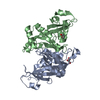
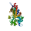
| Title | 2.45 Angstrom Crystal Structure of the Complex Between the Nuclear SnoRNA Decapping Nudix Hydrolase X29 and Manganese in the Presence of 7-methyl-GTP | ||||||
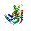
 Components Components | U8 snoRNA-binding protein X29 | ||||||
 Keywords Keywords | TRANSLATION / HYDROLASE / modified nudix hydrolase fold | ||||||
| Function / homology |  Function and homology information Function and homology informationinosine diphosphate phosphatase / sno(s)RNA catabolic process / dIDP phosphatase activity / dITP catabolic process / IDP phosphatase activity / RNA NAD+-cap (NAD+-forming) hydrolase activity / dITP diphosphatase activity / negative regulation of rRNA processing / phosphodiesterase decapping endonuclease activity / positive regulation of cell cycle process ...inosine diphosphate phosphatase / sno(s)RNA catabolic process / dIDP phosphatase activity / dITP catabolic process / IDP phosphatase activity / RNA NAD+-cap (NAD+-forming) hydrolase activity / dITP diphosphatase activity / negative regulation of rRNA processing / phosphodiesterase decapping endonuclease activity / positive regulation of cell cycle process / NAD-cap decapping / 5'-(N7-methylguanosine 5'-triphospho)-[mRNA] hydrolase / 5'-(N(7)-methylguanosine 5'-triphospho)-[mRNA] hydrolase activity / metalloexopeptidase activity / cobalt ion binding / snoRNA binding / mRNA catabolic process / manganese ion binding / nucleotide binding / mRNA binding / nucleolus / magnesium ion binding / protein homodimerization activity / nucleoplasm / nucleus / cytoplasm Similarity search - Function | ||||||
| Biological species | |||||||
| Method |  X-RAY DIFFRACTION / MOLECULAR REPLACEMENT / Resolution: 2.45 Å X-RAY DIFFRACTION / MOLECULAR REPLACEMENT / Resolution: 2.45 Å | ||||||
 Authors Authors | Scarsdale, J.N. / Peculis, B.A. / Wright, H.T. | ||||||
 Citation Citation | Journal: Structure / Year: 2006 Title: Crystal structures of U8 snoRNA decapping nudix hydrolase, X29, and its metal and cap complexes Authors: Scarsdale, J.N. / Peculis, B.A. / Wright, H.T. #1: Journal: Acta Crystallogr.,Sect.D / Year: 2004Title: Crystals of X29, a Xenopus Laevis U8 SnoRNA Binding Protein with Nuclear Decapping Activity Authors: Peculis, B.A. / Scarsdale, J.N. / Wright, H.T. #2: Journal: Mol.Cell / Year: 2004Title: Xenopus U8 SnoRNA Binding Protein is a Conserved Nuclear Decapping Enzyme Authors: Ghosh, T. / Peterson, B. / Tomasevic, N. / Peculis, B.A. | ||||||
| History |
|
- Structure visualization
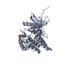
| Structure viewer | Molecule: MolmilJmol/JSmol |
|---|
- Downloads & links
-Download
| PDBx/mmCIF format | 2a8r.cif.gz | 92.1 KB | Display | PDBx/mmCIF format |
|---|---|---|---|---|
| PDB format | pdb2a8r.ent.gz | 68.5 KB | Display | PDB format |
| PDBx/mmJSON format | 2a8r.json.gz | Tree view | PDBx/mmJSON format | |
| Others |  Other downloads Other downloads |
-Validation report
| Arichive directory | https://data.pdbj.org/pub/pdb/validation_reports/a8/2a8rftp://data.pdbj.org/pub/pdb/validation_reports/a8/2a8r | HTTPS FTP |
|---|
-Related structure data
| Related structure data | 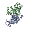 2a8pC 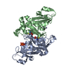 2a8qC 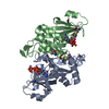 2a8sC 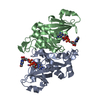 2a8tC  1u20S S: Starting model for refinement C: citing same article ( |
|---|---|
| Similar structure data |


-Links
 PDBj
PDBj


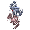
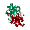
- Assembly
| Deposited unit | 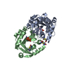
| ||||||||||
|---|---|---|---|---|---|---|---|---|---|---|---|
| 1 |
| ||||||||||
| Unit cell |
|
-Components
| #1: Protein | Mass: 24504.127 Da / Num. of mol.: 2 Source method: isolated from a genetically manipulated source Source: (gene. exp.)  References: UniProt: Q569R2, UniProt: Q6TEC1*PLUS, Hydrolases; Acting on acid anhydrides; In phosphorus-containing anhydrides #2: Chemical | ChemComp-MN /   Mass: 54.938 Da / Num. of mol.: 5 / Source method: obtained synthetically / Formula: Mn Mass: 54.938 Da / Num. of mol.: 5 / Source method: obtained synthetically / Formula: Mn#3: Chemical |   Mass: 175.959 Da / Num. of mol.: 2 / Source method: obtained synthetically / Formula: H2O7P2 Mass: 175.959 Da / Num. of mol.: 2 / Source method: obtained synthetically / Formula: H2O7P2#4: Water | ChemComp-HOH / |  Mass: 18.015 Da / Num. of mol.: 73 / Source method: isolated from a natural source / Formula: H2O Mass: 18.015 Da / Num. of mol.: 73 / Source method: isolated from a natural source / Formula: H2O |
|---|
-Experimental details
-Experiment
| Experiment | Method: X-RAY DIFFRACTION / Number of used crystals: 1 |
|---|
- Sample preparation
| Crystal | Density Matthews: 2.27 Å3/Da / Density % sol: 45.3 % |
|---|---|
| Crystal grow | Temperature: 293 K / Method: vapor diffusion, sitting drop / pH: 7.68 Details: 4-5 mg/ml X29, 0.025M HEPES pH 7.68, 3/75% PEG 6000, VAPOR DIFFUSION, SITTING DROP, temperature 293K |
-Data collection
| Diffraction | Mean temperature: 100 K | |||||||||||||||||||||||||||||||||||||||||||||||||||||||||||||||||||||||||||||
|---|---|---|---|---|---|---|---|---|---|---|---|---|---|---|---|---|---|---|---|---|---|---|---|---|---|---|---|---|---|---|---|---|---|---|---|---|---|---|---|---|---|---|---|---|---|---|---|---|---|---|---|---|---|---|---|---|---|---|---|---|---|---|---|---|---|---|---|---|---|---|---|---|---|---|---|---|---|---|
| Diffraction source | Source: ROTATING ANODE / Type: RIGAKU / Wavelength: 1.5418 Å | |||||||||||||||||||||||||||||||||||||||||||||||||||||||||||||||||||||||||||||
| Detector | Type: RIGAKU RAXIS IIC / Detector: IMAGE PLATE / Date: Apr 14, 2004 / Details: Osmic Confocal Optics | |||||||||||||||||||||||||||||||||||||||||||||||||||||||||||||||||||||||||||||
| Radiation | Monochromator: Osmic Confocal Optics / Protocol: SINGLE WAVELENGTH / Monochromatic (M) / Laue (L): M / Scattering type: x-ray | |||||||||||||||||||||||||||||||||||||||||||||||||||||||||||||||||||||||||||||
| Radiation wavelength | Wavelength: 1.5418 Å / Relative weight: 1 | |||||||||||||||||||||||||||||||||||||||||||||||||||||||||||||||||||||||||||||
| Reflection | Redundancy: 3.46 % / Av σ(I) over netI: 14.1 / Number: 754 / Rmerge(I) obs: 0.046 / Χ2: 0.99 / D res high: 2.45 Å / D res low: 23.51 Å / Num. obs: 17613 / % possible obs: 99.4 / Rejects: 460 | |||||||||||||||||||||||||||||||||||||||||||||||||||||||||||||||||||||||||||||
| Diffraction reflection shell |
| |||||||||||||||||||||||||||||||||||||||||||||||||||||||||||||||||||||||||||||
| Reflection | Resolution: 2.45→23.51 Å / Num. all: 17716 / Num. obs: 17613 / % possible obs: 99.4 % / Redundancy: 3.46 % / Rmerge(I) obs: 0.046 / Net I/σ(I): 14.1 | |||||||||||||||||||||||||||||||||||||||||||||||||||||||||||||||||||||||||||||
| Reflection shell | Resolution: 2.45→2.54 Å / Redundancy: 3.48 % / Rmerge(I) obs: 0.365 / Mean I/σ(I) obs: 3.1 / Num. unique all: 1727 / % possible all: 99.5 |
- Processing
| Software |
| |||||||||||||||||||||||||||||||||||||||||||||||||||||||||||||||||||||||||||||||||||||
|---|---|---|---|---|---|---|---|---|---|---|---|---|---|---|---|---|---|---|---|---|---|---|---|---|---|---|---|---|---|---|---|---|---|---|---|---|---|---|---|---|---|---|---|---|---|---|---|---|---|---|---|---|---|---|---|---|---|---|---|---|---|---|---|---|---|---|---|---|---|---|---|---|---|---|---|---|---|---|---|---|---|---|---|---|---|---|
| Refinement | Method to determine structure: MOLECULAR REPLACEMENT Starting model: PDB entry 1U20 Resolution: 2.45→23 Å / Cor.coef. Fo:Fc: 0.945 / Cor.coef. Fo:Fc free: 0.926 / SU B: 19.678 / SU ML: 0.217 / TLS residual ADP flag: LIKELY RESIDUAL Isotropic thermal model: TLS refinement followd by restrained refinement of individual B factors Cross valid method: THROUGHOUT / σ(F): 0 / ESU R: 0.507 / ESU R Free: 0.292 / Stereochemistry target values: MAXIMUM LIKELIHOOD Details: Simulated annealing via Torsion Angle Dynamics in CNS v1.0 was followed by refinement with REFMAC5,
| |||||||||||||||||||||||||||||||||||||||||||||||||||||||||||||||||||||||||||||||||||||
| Solvent computation | Ion probe radii: 0.8 Å / Shrinkage radii: 0.8 Å / VDW probe radii: 1.2 Å / Solvent model: BABINET MODEL WITH MASK | |||||||||||||||||||||||||||||||||||||||||||||||||||||||||||||||||||||||||||||||||||||
| Displacement parameters | Biso mean: 63.01 Å2
| |||||||||||||||||||||||||||||||||||||||||||||||||||||||||||||||||||||||||||||||||||||
| Refine analyze | Luzzati coordinate error obs: 0.448 Å | |||||||||||||||||||||||||||||||||||||||||||||||||||||||||||||||||||||||||||||||||||||
| Refinement step | Cycle: LAST / Resolution: 2.45→23 Å
| |||||||||||||||||||||||||||||||||||||||||||||||||||||||||||||||||||||||||||||||||||||
| Refine LS restraints |
| |||||||||||||||||||||||||||||||||||||||||||||||||||||||||||||||||||||||||||||||||||||
| LS refinement shell | Resolution: 2.45→2.513 Å / Total num. of bins used: 20
| |||||||||||||||||||||||||||||||||||||||||||||||||||||||||||||||||||||||||||||||||||||
| Refinement TLS params. | Method: refined / Refine-ID: X-RAY DIFFRACTION
| |||||||||||||||||||||||||||||||||||||||||||||||||||||||||||||||||||||||||||||||||||||
| Refinement TLS group | Refine-ID: X-RAY DIFFRACTION / Selection: ALL
|
