| 登録情報 | データベース: PDB / ID: 2a5s
|
|---|
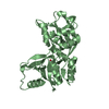
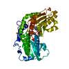
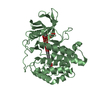
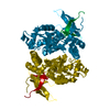
| タイトル | Crystal Structure Of The NR2A Ligand Binding Core In Complex With Glutamate |
|---|
 要素 要素 | N-methyl-D-aspartate receptor NMDAR2A subunit |
|---|
 キーワード キーワード | METAL TRANSPORT / MEMBRANE PROTEIN / Protein-ligand complex |
|---|
| 機能・相同性 |  機能・相同性情報 機能・相同性情報
regulation of response to alcohol / response to ammonium ion / neurotransmitter receptor transport, plasma membrane to endosome / receptor recycling / response to environmental enrichment / directional locomotion / Assembly and cell surface presentation of NMDA receptors / auditory behavior / response to other organism / response to hydrogen sulfide ...regulation of response to alcohol / response to ammonium ion / neurotransmitter receptor transport, plasma membrane to endosome / receptor recycling / response to environmental enrichment / directional locomotion / Assembly and cell surface presentation of NMDA receptors / auditory behavior / response to other organism / response to hydrogen sulfide / cellular response to magnesium ion / response to methylmercury / protein localization to postsynaptic membrane / serotonin metabolic process / regulation of ARF protein signal transduction / response to manganese ion / sleep / positive regulation of inhibitory postsynaptic potential / response to carbohydrate / cellular response to lipid / regulation of NMDA receptor activity / cellular response to dsRNA / dendritic spine organization / locomotion / response to amine / response to glycoside / Synaptic adhesion-like molecules / regulation of monoatomic cation transmembrane transport / NMDA glutamate receptor activity / voltage-gated monoatomic cation channel activity / NMDA selective glutamate receptor complex / glutamate binding / glutamate receptor signaling pathway / calcium ion transmembrane import into cytosol / spinal cord development / startle response / dopamine metabolic process / cellular response to zinc ion / parallel fiber to Purkinje cell synapse / response to lithium ion / regulation of postsynaptic membrane potential / monoatomic cation transmembrane transport / action potential / cellular response to glycine / modulation of excitatory postsynaptic potential / response to light stimulus / Unblocking of NMDA receptors, glutamate binding and activation / regulation of neuronal synaptic plasticity / positive regulation of protein targeting to membrane / glutamate receptor binding / conditioned place preference / multicellular organismal response to stress / neuron development / postsynaptic density, intracellular component / monoatomic cation channel activity / response to fungicide / positive regulation of synaptic transmission, glutamatergic / cellular response to manganese ion / glutamate-gated receptor activity / glutamate-gated calcium ion channel activity / cell adhesion molecule binding / sensory perception of pain / ionotropic glutamate receptor signaling pathway / neurogenesis / dendrite membrane / cytoplasmic vesicle membrane / ligand-gated monoatomic ion channel activity involved in regulation of presynaptic membrane potential / protein tyrosine kinase binding / positive regulation of excitatory postsynaptic potential / sodium ion transmembrane transport / response to amphetamine / learning / response to nicotine / synaptic membrane / hippocampus development / response to cocaine / cellular response to amino acid stimulus / synaptic transmission, glutamatergic / excitatory postsynaptic potential / regulation of membrane potential / transmitter-gated monoatomic ion channel activity involved in regulation of postsynaptic membrane potential / negative regulation of protein catabolic process / cerebral cortex development / visual learning / response to calcium ion / regulation of synaptic plasticity / regulation of long-term neuronal synaptic plasticity / response to wounding / response to lead ion / postsynaptic density membrane / cellular response to growth factor stimulus / modulation of chemical synaptic transmission / calcium ion transmembrane transport / calcium channel activity / memory / terminal bouton / calcium-dependent protein binding / synaptic vesicle / calcium ion transport / long-term synaptic potentiation類似検索 - 分子機能 Glutamate [NMDA] receptor, epsilon subunit, C-terminal / N-methyl D-aspartate receptor 2B3 C-terminus / Ionotropic glutamate receptor, metazoa / Ligated ion channel L-glutamate- and glycine-binding site / Ligand-gated ion channel / Ionotropic glutamate receptor, L-glutamate and glycine-binding domain / Ligated ion channel L-glutamate- and glycine-binding site / : / Ionotropic glutamate receptor / Eukaryotic homologues of bacterial periplasmic substrate binding proteins. ...Glutamate [NMDA] receptor, epsilon subunit, C-terminal / N-methyl D-aspartate receptor 2B3 C-terminus / Ionotropic glutamate receptor, metazoa / Ligated ion channel L-glutamate- and glycine-binding site / Ligand-gated ion channel / Ionotropic glutamate receptor, L-glutamate and glycine-binding domain / Ligated ion channel L-glutamate- and glycine-binding site / : / Ionotropic glutamate receptor / Eukaryotic homologues of bacterial periplasmic substrate binding proteins. / Periplasmic binding protein-like II / Receptor, ligand binding region / Receptor family ligand binding region / D-Maltodextrin-Binding Protein; domain 2 / Periplasmic binding protein-like I / 3-Layer(aba) Sandwich / Alpha Beta類似検索 - ドメイン・相同性 GLUTAMIC ACID / : / Glutamate receptor ionotropic, NMDA 2A類似検索 - 構成要素 |
|---|
| 生物種 |   Rattus norvegicus (ドブネズミ) Rattus norvegicus (ドブネズミ) |
|---|
| 手法 |  X線回折 / シンクロトロン / Combination of molecular replacement, 単波長異常分散 / 解像度: 1.7 Å X線回折 / シンクロトロン / Combination of molecular replacement, 単波長異常分散 / 解像度: 1.7 Å |
|---|
 データ登録者 データ登録者 | Furukawa, H. / Singh, S.K. / Mancusso, R. / Gouaux, E. |
|---|
 引用 引用 | ジャーナル: Nature / 年: 2005
タイトル: Subunit arrangement and function in NMDA receptors
著者: Furukawa, H. / Singh, S.K. / Mancusso, R. / Gouaux, E. |
|---|
| 履歴 | | 登録 | 2005年6月30日 | 登録サイト: RCSB / 処理サイト: RCSB |
|---|
| 改定 1.0 | 2005年11月15日 | Provider: repository / タイプ: Initial release |
|---|
| 改定 1.1 | 2008年4月30日 | Group: Version format compliance |
|---|
| 改定 1.2 | 2011年7月13日 | Group: Version format compliance |
|---|
| 改定 1.3 | 2017年7月26日 | Group: Refinement description / Source and taxonomy / カテゴリ: entity_src_gen / software |
|---|
| 改定 1.4 | 2024年10月30日 | Group: Data collection / Database references ...Data collection / Database references / Derived calculations / Structure summary
カテゴリ: chem_comp_atom / chem_comp_bond ...chem_comp_atom / chem_comp_bond / database_2 / pdbx_entry_details / pdbx_modification_feature / struct_site
Item: _database_2.pdbx_DOI / _database_2.pdbx_database_accession ..._database_2.pdbx_DOI / _database_2.pdbx_database_accession / _struct_site.pdbx_auth_asym_id / _struct_site.pdbx_auth_comp_id / _struct_site.pdbx_auth_seq_id |
|---|
|
|---|
 ムービー
ムービー コントローラー


 データを開く
 基本情報
基本情報 構造の表示
構造の表示 ダウンロードとリンク
 その他のダウンロード


 PDBj
PDBj


 集合体
集合体


 タイプ: L-peptide linking / 分子量: 147.129 Da / 分子数: 1 / 由来タイプ: 合成 / 式: C5H9NO4
 分子量: 18.015 Da / 分子数: 316 / 由来タイプ: 天然 / 式: H2O
 試料調製
試料調製 / ビームライン: X4A / 波長: 0.9793 Å
 解析
解析