 Movie
Movie Controller

[English] 日本語
 Yorodumi
- PDB-2a1o: Crystal structure of ferrous dioxygen complex of T252A cytochrome... -
+ Open data

- Basic information
| Entry | Database: PDB / ID: 2a1o | ||||||
|---|---|---|---|---|---|---|---|
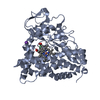
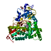
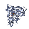
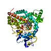
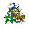
| Title | Crystal structure of ferrous dioxygen complex of T252A cytochrome P450cam | ||||||
 Components Components | Cytochrome P450-cam | ||||||
 Keywords Keywords | OXIDOREDUCTASE / cytochrome P450 / CYP / dioxygen complex / oxy | ||||||
| Function / homology |  Function and homology information Function and homology informationcamphor 5-monooxygenase / camphor 5-monooxygenase activity / (+)-camphor catabolic process / iron ion binding / heme binding / cytoplasm Similarity search - Function | ||||||
| Biological species |  Pseudomonas putida (bacteria) Pseudomonas putida (bacteria) | ||||||
| Method |  X-RAY DIFFRACTION / SYNCHROTRON / MOLECULAR REPLACEMENT / Resolution: 1.55 Å X-RAY DIFFRACTION / SYNCHROTRON / MOLECULAR REPLACEMENT / Resolution: 1.55 Å | ||||||
 Authors Authors | Nagano, S. / Poulos, T.L. | ||||||
 Citation Citation | Journal: J.Biol.Chem. / Year: 2005 Title: Crystallographic study on the dioxygen complex of wild-type and mutant cytochrome P450cam. Implications for the dioxygen activation mechanism Authors: Nagano, S. / Poulos, T.L. #1: Journal: J.Biol.Chem. / Year: 2005Title: Crystal Structures of the Ferrous Dioxygen Complex of Wild-type Cytochrome P450eryF and Its Mutants, A245S and A245T: INVESTIGATION OF THE PROTON TRANSFER SYSTEM IN P450eryF Authors: Nagano, S. / Cupp-Vickery, J.R. / Poulos, T.L. | ||||||
| History |
|
- Structure visualization
| Structure viewer | Molecule: MolmilJmol/JSmol |
|---|
- Downloads & links
-Download
| PDBx/mmCIF format | 2a1o.cif.gz | 187.1 KB | Display | PDBx/mmCIF format |
|---|---|---|---|---|
| PDB format | pdb2a1o.ent.gz | 146.5 KB | Display | PDB format |
| PDBx/mmJSON format | 2a1o.json.gz | Tree view | PDBx/mmJSON format | |
| Others |  Other downloads Other downloads |
-Validation report
| Arichive directory | https://data.pdbj.org/pub/pdb/validation_reports/a1/2a1oftp://data.pdbj.org/pub/pdb/validation_reports/a1/2a1o | HTTPS FTP |
|---|
-Related structure data
| Related structure data |  2a1mC  2a1nC  1dz4S C: citing same article ( S: Starting model for refinement |
|---|---|
| Similar structure data |


-Links
 PDBj
PDBj


- Assembly
| Deposited unit | 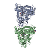
| ||||||||
|---|---|---|---|---|---|---|---|---|---|
| 1 | 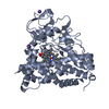
| ||||||||
| 2 | 
| ||||||||
| Unit cell |
|
-Components
-Protein , 1 types, 2 molecules AB
| #1: Protein | Mass: 46657.988 Da / Num. of mol.: 2 / Mutation: T252A/C334A Source method: isolated from a genetically manipulated source Source: (gene. exp.) Pseudomonas putida (bacteria) / Gene: CAMC / Plasmid: pUC19 / Production host: |
|---|
-Non-polymers , 6 types, 516 molecules 


| #2: Chemical |  Mass: 39.098 Da / Num. of mol.: 3 / Source method: obtained synthetically / Formula: K Mass: 39.098 Da / Num. of mol.: 3 / Source method: obtained synthetically / Formula: K#3: Chemical |  Mass: 616.487 Da / Num. of mol.: 2 / Source method: obtained synthetically / Formula: C34H32FeN4O4 Mass: 616.487 Da / Num. of mol.: 2 / Source method: obtained synthetically / Formula: C34H32FeN4O4#4: Chemical |  Mass: 31.999 Da / Num. of mol.: 2 / Source method: obtained synthetically / Formula: O2 Mass: 31.999 Da / Num. of mol.: 2 / Source method: obtained synthetically / Formula: O2#5: Chemical |  Mass: 152.233 Da / Num. of mol.: 2 / Source method: obtained synthetically / Formula: C10H16O Mass: 152.233 Da / Num. of mol.: 2 / Source method: obtained synthetically / Formula: C10H16O#6: Chemical | ChemComp-TRS / |  Mass: 122.143 Da / Num. of mol.: 1 / Source method: obtained synthetically / Formula: C4H12NO3 / Comment: pH buffer*YM Mass: 122.143 Da / Num. of mol.: 1 / Source method: obtained synthetically / Formula: C4H12NO3 / Comment: pH buffer*YM#7: Water | ChemComp-HOH / | Mass: 18.015 Da / Num. of mol.: 506 / Source method: isolated from a natural source / Formula: H2O |
|---|
-Experimental details
-Experiment
| Experiment | Method: X-RAY DIFFRACTION / Number of used crystals: 1 |
|---|
- Sample preparation
| Crystal | Density Matthews: 2.1 Å3/Da / Density % sol: 43 % |
|---|---|
| Crystal grow | Temperature: 288 K / Method: vapor diffusion, sitting drop / pH: 7.4 Details: PEG 4000, potassium chloride, Tris-HCl, D-camphor, pH 7.4, VAPOR DIFFUSION, SITTING DROP, temperature 288K |
-Data collection
| Diffraction | Mean temperature: 100 K |
|---|---|
| Diffraction source | Source: SYNCHROTRON / Site: SSRL  / Beamline: BL9-2 / Wavelength: 0.92 Å / Beamline: BL9-2 / Wavelength: 0.92 Å |
| Detector | Date: Mar 31, 2005 |
| Radiation | Protocol: SINGLE WAVELENGTH / Monochromatic (M) / Laue (L): M / Scattering type: x-ray |
| Radiation wavelength | Wavelength: 0.92 Å / Relative weight: 1 |
| Reflection | Resolution: 1.55→41.07 Å / Num. obs: 108552 / % possible obs: 95.3 % / Observed criterion σ(I): -3 / Biso Wilson estimate: 17.6 Å2 / Net I/σ(I): 27.8 |
| Reflection shell | Resolution: 1.55→1.58 Å / Mean I/σ(I) obs: 2.3 |
- Processing
| Software | Name: CNS / Version: 1.1 / Classification: refinement | ||||||||||||||||||||||||||||||||||||||||||||||||||||||||||||||||||||||||||||||||
|---|---|---|---|---|---|---|---|---|---|---|---|---|---|---|---|---|---|---|---|---|---|---|---|---|---|---|---|---|---|---|---|---|---|---|---|---|---|---|---|---|---|---|---|---|---|---|---|---|---|---|---|---|---|---|---|---|---|---|---|---|---|---|---|---|---|---|---|---|---|---|---|---|---|---|---|---|---|---|---|---|---|
| Refinement | Method to determine structure: MOLECULAR REPLACEMENT Starting model: PDB ENTRY 1DZ4 Resolution: 1.55→41.07 Å / Rfactor Rfree error: 0.003 / Data cutoff high absF: 258087.24 / Data cutoff low absF: 0 / Isotropic thermal model: RESTRAINED / Cross valid method: THROUGHOUT / σ(F): 0
| ||||||||||||||||||||||||||||||||||||||||||||||||||||||||||||||||||||||||||||||||
| Solvent computation | Solvent model: FLAT MODEL / Bsol: 45.5631 Å2 / ksol: 0.360128 e/Å3 | ||||||||||||||||||||||||||||||||||||||||||||||||||||||||||||||||||||||||||||||||
| Displacement parameters | Biso mean: 19.9 Å2
| ||||||||||||||||||||||||||||||||||||||||||||||||||||||||||||||||||||||||||||||||
| Refine analyze |
| ||||||||||||||||||||||||||||||||||||||||||||||||||||||||||||||||||||||||||||||||
| Refinement step | Cycle: LAST / Resolution: 1.55→41.07 Å
| ||||||||||||||||||||||||||||||||||||||||||||||||||||||||||||||||||||||||||||||||
| Refine LS restraints |
| ||||||||||||||||||||||||||||||||||||||||||||||||||||||||||||||||||||||||||||||||
| LS refinement shell | Resolution: 1.55→1.58 Å / Rfactor Rfree error: 0.023 / Total num. of bins used: 20
| ||||||||||||||||||||||||||||||||||||||||||||||||||||||||||||||||||||||||||||||||
| Xplor file |
|
