 Movie
Movie Controller

[English] 日本語
 Yorodumi
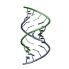
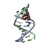
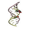
- PDB-205d: STRUCTURE OF AN RNA DOUBLE HELIX INCLUDING URACIL-URACIL BASE PAI... -
+ Open data

- Basic information
| Entry | Database: PDB / ID: 205d | ||||||||||||||||||
|---|---|---|---|---|---|---|---|---|---|---|---|---|---|---|---|---|---|---|---|
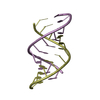
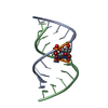
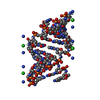
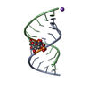
| Title | STRUCTURE OF AN RNA DOUBLE HELIX INCLUDING URACIL-URACIL BASE PAIRS IN AN INTERNAL LOOP | ||||||||||||||||||
 Components Components | RNA (5'-R(* Keywords KeywordsRNA / A-RNA / DOUBLE HELIX / INTERNAL LOOP / MISMATCHED | Function / homology | RNA / RNA (> 10) |  Function and homology information Function and homology informationMethod |  X-RAY DIFFRACTION / SYNCHROTRON / Resolution: 2.64 Å X-RAY DIFFRACTION / SYNCHROTRON / Resolution: 2.64 Å  Authors AuthorsBaeyens, K.J. / De Bondt, H.L. / Holbrook, S.R. |  CitationJournal: Nat.Struct.Biol. / Year: 1995 CitationJournal: Nat.Struct.Biol. / Year: 1995Title: Structure of an RNA double helix including uracil-uracil base pairs in an internal loop. Authors: Baeyens, K.J. / De Bondt, H.L. / Holbrook, S.R. History |
|
- Structure visualization
| Structure viewer | Molecule: MolmilJmol/JSmol |
|---|
- Downloads & links
-Download
| PDBx/mmCIF format | 205d.cif.gz | 24.1 KB | Display | PDBx/mmCIF format |
|---|---|---|---|---|
| PDB format | pdb205d.ent.gz | 15.3 KB | Display | PDB format |
| PDBx/mmJSON format | 205d.json.gz | Tree view | PDBx/mmJSON format | |
| Others |  Other downloads Other downloads |
-Validation report
| Arichive directory | https://data.pdbj.org/pub/pdb/validation_reports/05/205dftp://data.pdbj.org/pub/pdb/validation_reports/05/205d | HTTPS FTP |
|---|
-Related structure data
| Similar structure data |
|---|


-Links
 PDBj
PDBj


- Assembly
| Deposited unit | 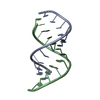
| ||||||||
|---|---|---|---|---|---|---|---|---|---|
| 1 |
| ||||||||
| Unit cell |
|
-Components
| #1: RNA chain | Mass: 3805.280 Da / Num. of mol.: 2 Source method: isolated from a genetically manipulated source #2: Water | ChemComp-HOH / |  Mass: 18.015 Da / Num. of mol.: 39 / Source method: isolated from a natural source / Formula: H2O Mass: 18.015 Da / Num. of mol.: 39 / Source method: isolated from a natural source / Formula: H2O |
|---|
-Experimental details
-Experiment
| Experiment | Method: X-RAY DIFFRACTION |
|---|
- Sample preparation
| Crystal | Density Matthews: 2.95 Å3/Da / Density % sol: 57 % | ||||||||||||||||||||||||||||||||||||||||||||||||||||||
|---|---|---|---|---|---|---|---|---|---|---|---|---|---|---|---|---|---|---|---|---|---|---|---|---|---|---|---|---|---|---|---|---|---|---|---|---|---|---|---|---|---|---|---|---|---|---|---|---|---|---|---|---|---|---|---|
| Crystal grow | Method: vapor diffusion, hanging drop / pH: 5.6 / Details: pH 5.60, VAPOR DIFFUSION, HANGING DROP | ||||||||||||||||||||||||||||||||||||||||||||||||||||||
| Components of the solutions |
| ||||||||||||||||||||||||||||||||||||||||||||||||||||||
| Crystal | *PLUS | ||||||||||||||||||||||||||||||||||||||||||||||||||||||
| Crystal grow | *PLUS pH: 5.6 | ||||||||||||||||||||||||||||||||||||||||||||||||||||||
| Components of the solutions | *PLUS
|
-Data collection
| Diffraction source | Source: SYNCHROTRON / Site: SSRL  / Type: SSRL / Type: SSRL |
|---|---|
| Detector | Type: MARRESEARCH / Detector: IMAGE PLATE / Date: Jul 1, 1993 |
| Radiation | Monochromatic (M) / Laue (L): M / Scattering type: x-ray |
| Radiation wavelength | Relative weight: 1 |
| Reflection | Highest resolution: 2.64 Å / Num. obs: 2415 / % possible obs: 90.1 % / Observed criterion σ(I): 2.6 / Rmerge(I) obs: 0.108 |
| Reflection | *PLUS Highest resolution: 2.64 Å / % possible obs: 90.1 % / Observed criterion σ(I): 2.6 |
- Processing
| Software | Name: X-PLOR / Classification: refinement | ||||||||||||||||||||||||||||||||||||||||||||||||||||||||||||
|---|---|---|---|---|---|---|---|---|---|---|---|---|---|---|---|---|---|---|---|---|---|---|---|---|---|---|---|---|---|---|---|---|---|---|---|---|---|---|---|---|---|---|---|---|---|---|---|---|---|---|---|---|---|---|---|---|---|---|---|---|---|
| Refinement | Resolution: 2.64→8 Å / σ(F): 2 /
| ||||||||||||||||||||||||||||||||||||||||||||||||||||||||||||
| Refinement step | Cycle: LAST / Resolution: 2.64→8 Å
| ||||||||||||||||||||||||||||||||||||||||||||||||||||||||||||
| Refine LS restraints |
| ||||||||||||||||||||||||||||||||||||||||||||||||||||||||||||
| Software | *PLUS Version: 3.1 / Classification: refinement | ||||||||||||||||||||||||||||||||||||||||||||||||||||||||||||
| Refinement | *PLUS Highest resolution: 2.64 Å / Lowest resolution: 8 Å / σ(F): 2 | ||||||||||||||||||||||||||||||||||||||||||||||||||||||||||||
| Solvent computation | *PLUS | ||||||||||||||||||||||||||||||||||||||||||||||||||||||||||||
| Displacement parameters | *PLUS |
