 Movie
Movie Controller

[English] 日本語
 Yorodumi
- PDB-1zlh: Crystal structure of the tick carboxypeptidase inhibitor in compl... -
+ Open data

- Basic information
| Entry | Database: PDB / ID: 1zlh | ||||||
|---|---|---|---|---|---|---|---|
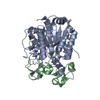
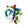
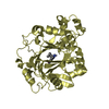
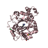
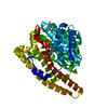
| Title | Crystal structure of the tick carboxypeptidase inhibitor in complex with bovine carboxypeptidase A | ||||||
 Components Components |
| ||||||
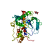
 Keywords Keywords | HYDROLASE/HYDROLASE INHIBITOR / inhibitor-metallocarboxypeptidase complex / beta-defensin fold (TCI) / eight-stranded twisted beta-sheet surrounded by eight alpha-helices (CPA) / HYDROLASE-HYDROLASE INHIBITOR COMPLEX | ||||||
| Function / homology |  Function and homology information Function and homology informationcarboxypeptidase A / acquisition of nutrients from host / metalloendopeptidase inhibitor activity / leukotriene metabolic process / metallocarboxypeptidase activity / enzyme inhibitor activity / toxin activity / proteolysis / : / extracellular region / zinc ion binding Similarity search - Function | ||||||
| Biological species |   Rhipicephalus bursa (arthropod) Rhipicephalus bursa (arthropod) | ||||||
| Method |  X-RAY DIFFRACTION / MIR / Resolution: 1.7 Å X-RAY DIFFRACTION / MIR / Resolution: 1.7 Å | ||||||
 Authors Authors | Arolas, J.L. / Popowicz, G.M. / Lorenzo, J. / Sommerhoff, C.P. / Huber, R. / Aviles, F.X. / Holak, T.A. | ||||||
 Citation Citation | Journal: J.Mol.Biol. / Year: 2005 Title: The Three-Dimensional Structures of Tick Carboxypeptidase Inhibitor in Complex with A/B Carboxypeptidases Reveal a Novel Double-headed Binding Mode Authors: Arolas, J.L. / Popowicz, G.M. / Lorenzo, J. / Sommerhoff, C.P. / Huber, R. / Aviles, F.X. / Holak, T.A. #1: Journal: J.Biol.Chem. / Year: 2005 Title: A carboxypeptidase inhibitor from the tick Rhipicephalus bursa: isolation, cDNA cloning, recombinant expression, and characterization Authors: Arolas, J.L. / Lorenzo, J. / Rovira, A. / Castella, J. / Aviles, F.X. / Sommerhoff, C.P. | ||||||
| History |
|
- Structure visualization
| Structure viewer | Molecule: MolmilJmol/JSmol |
|---|
- Downloads & links
-Download
| PDBx/mmCIF format | 1zlh.cif.gz | 163 KB | Display | PDBx/mmCIF format |
|---|---|---|---|---|
| PDB format | pdb1zlh.ent.gz | 126.6 KB | Display | PDB format |
| PDBx/mmJSON format | 1zlh.json.gz | Tree view | PDBx/mmJSON format | |
| Others |  Other downloads Other downloads |
-Validation report
| Arichive directory | https://data.pdbj.org/pub/pdb/validation_reports/zl/1zlhftp://data.pdbj.org/pub/pdb/validation_reports/zl/1zlh | HTTPS FTP |
|---|
-Related structure data


-Links
 PDBj
PDBj
- Assembly
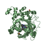
| Deposited unit | 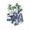
| ||||||||
|---|---|---|---|---|---|---|---|---|---|
| 1 |
| ||||||||
| Unit cell |
|
-Components
| #1: Protein | Mass: 34721.750 Da / Num. of mol.: 1 Source method: isolated from a genetically manipulated source Source: (gene. exp.)  Pichia pastoris (fungus) / Strain (production host): KM71 / References: UniProt: P00730, carboxypeptidase A Pichia pastoris (fungus) / Strain (production host): KM71 / References: UniProt: P00730, carboxypeptidase A | ||||
|---|---|---|---|---|---|
| #2: Protein | Mass: 7945.104 Da / Num. of mol.: 1 Source method: isolated from a genetically manipulated source Source: (gene. exp.) Rhipicephalus bursa (arthropod) / Plasmid: pBAT4 / Species (production host): Escherichia coli / Production host:  | ||||
| #3: Chemical | ChemComp-ZN /   Mass: 65.409 Da / Num. of mol.: 5 / Source method: obtained synthetically / Formula: Zn Mass: 65.409 Da / Num. of mol.: 5 / Source method: obtained synthetically / Formula: Zn#4: Water | ChemComp-HOH / |  Mass: 18.015 Da / Num. of mol.: 379 / Source method: isolated from a natural source / Formula: H2O Mass: 18.015 Da / Num. of mol.: 379 / Source method: isolated from a natural source / Formula: H2OHas protein modification | Y | |
-Experimental details
-Experiment
| Experiment | Method: X-RAY DIFFRACTION / Number of used crystals: 1 |
|---|
- Sample preparation
| Crystal | Density Matthews: 2.1 Å3/Da / Density % sol: 40.8 % |
|---|---|
| Crystal grow | Temperature: 293 K / Method: vapor diffusion, hanging drop / pH: 6.5 Details: 0.2M sodium cacodylate, 0.2M zinc acetate dihydrate, 7%(w/v) PEG 8000, 10%(w/v) dried dioxan, pH 6.5, VAPOR DIFFUSION, HANGING DROP, temperature 293.0K |
-Data collection
| Diffraction | Mean temperature: 100 K |
|---|---|
| Diffraction source | Source: ROTATING ANODE / Type: RIGAKU / Wavelength: 1.5418 |
| Detector | Type: MAR scanner 345 mm plate / Detector: IMAGE PLATE / Date: Feb 7, 2004 / Details: mirror |
| Radiation | Monochromator: mirror / Protocol: SINGLE WAVELENGTH / Monochromatic (M) / Laue (L): M / Scattering type: x-ray |
| Radiation wavelength | Wavelength: 1.5418 Å / Relative weight: 1 |
| Reflection | Resolution: 1.67→30 Å / Num. all: 39265 / Num. obs: 34176 / % possible obs: 87 % / Observed criterion σ(F): 2 / Observed criterion σ(I): 2 / Redundancy: 3.7 % / Rmerge(I) obs: 0.06 |
| Reflection shell | Resolution: 1.67→1.8 Å / Rmerge(I) obs: 0.134 / Num. unique all: 3786 / % possible all: 61.5 |
- Processing
| Software |
| ||||||||||||||||||||||||||||||||||||||||||||||||||||||||||||||||||||||||||||||||||||||||||||||||||||||||||||||||||||||||||||||||||||||||||||||||||||||||||||||||||||||||||
|---|---|---|---|---|---|---|---|---|---|---|---|---|---|---|---|---|---|---|---|---|---|---|---|---|---|---|---|---|---|---|---|---|---|---|---|---|---|---|---|---|---|---|---|---|---|---|---|---|---|---|---|---|---|---|---|---|---|---|---|---|---|---|---|---|---|---|---|---|---|---|---|---|---|---|---|---|---|---|---|---|---|---|---|---|---|---|---|---|---|---|---|---|---|---|---|---|---|---|---|---|---|---|---|---|---|---|---|---|---|---|---|---|---|---|---|---|---|---|---|---|---|---|---|---|---|---|---|---|---|---|---|---|---|---|---|---|---|---|---|---|---|---|---|---|---|---|---|---|---|---|---|---|---|---|---|---|---|---|---|---|---|---|---|---|---|---|---|---|---|---|---|
| Refinement | Method to determine structure: MIR / Resolution: 1.7→55.9 Å / Cor.coef. Fo:Fc: 0.96 / Cor.coef. Fo:Fc free: 0.944 / SU B: 1.988 / SU ML: 0.065 / Cross valid method: THROUGHOUT / σ(F): 2 / ESU R: 0.225 / ESU R Free: 0.102 / Stereochemistry target values: MAXIMUM LIKELIHOOD / Details: HYDROGENS HAVE BEEN ADDED IN THE RIDING POSITIONS
| ||||||||||||||||||||||||||||||||||||||||||||||||||||||||||||||||||||||||||||||||||||||||||||||||||||||||||||||||||||||||||||||||||||||||||||||||||||||||||||||||||||||||||
| Solvent computation | Ion probe radii: 0.8 Å / Shrinkage radii: 0.8 Å / VDW probe radii: 1.4 Å / Solvent model: BABINET MODEL WITH MASK | ||||||||||||||||||||||||||||||||||||||||||||||||||||||||||||||||||||||||||||||||||||||||||||||||||||||||||||||||||||||||||||||||||||||||||||||||||||||||||||||||||||||||||
| Displacement parameters | Biso mean: 12.551 Å2
| ||||||||||||||||||||||||||||||||||||||||||||||||||||||||||||||||||||||||||||||||||||||||||||||||||||||||||||||||||||||||||||||||||||||||||||||||||||||||||||||||||||||||||
| Refinement step | Cycle: LAST / Resolution: 1.7→55.9 Å
| ||||||||||||||||||||||||||||||||||||||||||||||||||||||||||||||||||||||||||||||||||||||||||||||||||||||||||||||||||||||||||||||||||||||||||||||||||||||||||||||||||||||||||
| Refine LS restraints |
| ||||||||||||||||||||||||||||||||||||||||||||||||||||||||||||||||||||||||||||||||||||||||||||||||||||||||||||||||||||||||||||||||||||||||||||||||||||||||||||||||||||||||||
| LS refinement shell | Resolution: 1.7→1.744 Å / Total num. of bins used: 20 /
|
