| 登録情報 | データベース: PDB / ID: 1zjh
|
|---|
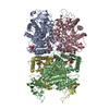
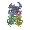
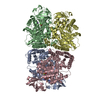
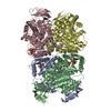
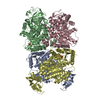
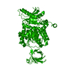
| タイトル | Structure of human muscle pyruvate kinase (PKM2) |
|---|
 要素 要素 | Pyruvate kinase, isozymes M1/M2 |
|---|
 キーワード キーワード | TRANSFERASE / muscpyruvate kinase / muscle isozyme / allosteric regulation / tranferase / Structural Genomics / Structural Genomics Consortium / SGC |
|---|
| 機能・相同性 |  機能・相同性情報 機能・相同性情報
pyruvate kinase / pyruvate kinase activity / histone H3T11 kinase activity / programmed cell death / Pyruvate metabolism / positive regulation of cytoplasmic translation / Glycolysis / canonical glycolysis / positive regulation of sprouting angiogenesis / potassium ion binding ...pyruvate kinase / pyruvate kinase activity / histone H3T11 kinase activity / programmed cell death / Pyruvate metabolism / positive regulation of cytoplasmic translation / Glycolysis / canonical glycolysis / positive regulation of sprouting angiogenesis / potassium ion binding / rough endoplasmic reticulum / Regulation of pyruvate metabolism / glycolytic process / non-specific protein-tyrosine kinase / non-membrane spanning protein tyrosine kinase activity / Negative Regulation of CDH1 Gene Transcription / cellular response to insulin stimulus / MHC class II protein complex binding / extracellular vesicle / sperm principal piece / protein tyrosine kinase activity / secretory granule lumen / vesicle / ficolin-1-rich granule lumen / transcription coactivator activity / non-specific serine/threonine protein kinase / cadherin binding / protein serine/threonine kinase activity / mRNA binding / Neutrophil degranulation / magnesium ion binding / protein homodimerization activity / positive regulation of transcription by RNA polymerase II / mitochondrion / RNA binding / extracellular exosome / extracellular region / nucleoplasm / ATP binding / nucleus / cytoplasm / cytosol類似検索 - 分子機能 PK beta-barrel domain-like / M1 Pyruvate Kinase; Domain 3 / Pyruvate kinase, C-terminal domain / Pyruvate Kinase; Chain: A, domain 1 / Pyruvate kinase, active site / Pyruvate kinase active site signature. / Pyruvate kinase / Pyruvate kinase, barrel / Pyruvate kinase, insert domain superfamily / Pyruvate kinase, barrel domain ...PK beta-barrel domain-like / M1 Pyruvate Kinase; Domain 3 / Pyruvate kinase, C-terminal domain / Pyruvate Kinase; Chain: A, domain 1 / Pyruvate kinase, active site / Pyruvate kinase active site signature. / Pyruvate kinase / Pyruvate kinase, barrel / Pyruvate kinase, insert domain superfamily / Pyruvate kinase, barrel domain / Pyruvate kinase, C-terminal / Pyruvate kinase, C-terminal domain superfamily / Pyruvate kinase, alpha/beta domain / Pyruvate kinase-like, insert domain superfamily / Phosphoenolpyruvate-binding domains / Pyruvate kinase-like domain superfamily / Pyruvate/Phosphoenolpyruvate kinase-like domain superfamily / TIM Barrel / Alpha-Beta Barrel / Beta Barrel / 3-Layer(aba) Sandwich / Mainly Beta / Alpha Beta類似検索 - ドメイン・相同性 |
|---|
| 生物種 |  Homo sapiens (ヒト) Homo sapiens (ヒト) |
|---|
| 手法 |  X線回折 / 分子置換 / 解像度: 2.2 Å X線回折 / 分子置換 / 解像度: 2.2 Å |
|---|
 データ登録者 データ登録者 | Choe, J. / Atanassova, A. / Arrowsmith, C. / Edwards, A. / Sundstrom, M. / Bochkarev, A. / Park, H. / Structural Genomics Consortium (SGC) |
|---|
 引用 引用 | ジャーナル: TO BE PUBLISHED
タイトル: Structure of human muscle pyruvate kinase (PKM2).
著者: Atanassova, A. / Choe, J. / Arrowsmith, C. / Edwards, A. / Sundstrom, M. / Bochkarev, A. / Park, H. |
|---|
| 履歴 | | 登録 | 2005年4月28日 | 登録サイト: RCSB / 処理サイト: RCSB |
|---|
| 改定 1.0 | 2005年5月17日 | Provider: repository / タイプ: Initial release |
|---|
| 改定 1.1 | 2008年4月30日 | Group: Version format compliance |
|---|
| 改定 1.2 | 2011年7月13日 | Group: Derived calculations / Version format compliance |
|---|
| 改定 1.3 | 2017年10月11日 | Group: Refinement description / カテゴリ: software / Item: _software.classification / _software.name |
|---|
| 改定 1.4 | 2023年8月23日 | Group: Data collection / Database references / Refinement description
カテゴリ: chem_comp_atom / chem_comp_bond ...chem_comp_atom / chem_comp_bond / database_2 / pdbx_initial_refinement_model / struct_ref_seq_dif
Item: _database_2.pdbx_DOI / _database_2.pdbx_database_accession / _struct_ref_seq_dif.details |
|---|
|
|---|
 ムービー
ムービー コントローラー


 データを開く
 基本情報
基本情報 構造の表示
構造の表示 ダウンロードとリンク
 その他のダウンロード
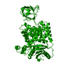


 PDBj
PDBj

 集合体
集合体

 分子量: 18.015 Da / 分子数: 122 / 由来タイプ: 天然 / 式: H2O
 試料調製
試料調製 解析
解析