 Movie
Movie Controller


+ Open data

- Basic information
| Entry | Database: PDB / ID: 1zhq | ||||||
|---|---|---|---|---|---|---|---|
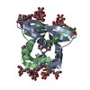
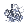
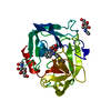
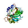
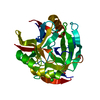
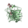
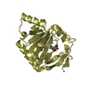
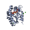
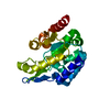
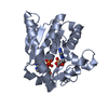
| Title | Crystal structure of apo MVL | ||||||
 Components Components | mannan-binding lectin | ||||||
 Keywords Keywords | SUGAR BINDING PROTEIN / HIV-1 / MVL / cyanobacteria / carbohydrate binding | ||||||
| Function / homology |  Function and homology information Function and homology informationregulation of defense response to virus / hydrolase activity, acting on glycosyl bonds / Hydrolases; Glycosylases; Glycosidases, i.e. enzymes that hydrolyse O- and S-glycosyl compounds / carbohydrate binding / cytoplasm Similarity search - Function | ||||||
| Biological species |  Microcystis viridis (bacteria) Microcystis viridis (bacteria) | ||||||
| Method |  X-RAY DIFFRACTION / MIR / Resolution: 1.9 Å X-RAY DIFFRACTION / MIR / Resolution: 1.9 Å | ||||||
 Authors Authors | Williams, D.C. / Lee, J.Y. / Cai, M. / Bewley, C.A. / Clore, G.M. | ||||||
 Citation Citation | Journal: J.Biol.Chem. / Year: 2005 Title: Crystal Structures of the HIV-1 Inhibitory Cyanobacterial Protein MVL Free and Bound to Man3GlcNAc2: STRUCTURAL BASIS FOR SPECIFICITY AND HIGH-AFFINITY BINDING TO THE CORE PENTASACCHARIDE FROM ...Title: Crystal Structures of the HIV-1 Inhibitory Cyanobacterial Protein MVL Free and Bound to Man3GlcNAc2: STRUCTURAL BASIS FOR SPECIFICITY AND HIGH-AFFINITY BINDING TO THE CORE PENTASACCHARIDE FROM N-LINKED OLIGOMANNOSIDE. Authors: Williams, D.C. / Lee, J.Y. / Cai, M. / Bewley, C.A. / Clore, G.M. | ||||||
| History |
|
- Structure visualization
| Structure viewer | Molecule: MolmilJmol/JSmol |
|---|
- Downloads & links
-Download
| PDBx/mmCIF format | 1zhq.cif.gz | 198.1 KB | Display | PDBx/mmCIF format |
|---|---|---|---|---|
| PDB format | pdb1zhq.ent.gz | 158.8 KB | Display | PDB format |
| PDBx/mmJSON format | 1zhq.json.gz | Tree view | PDBx/mmJSON format | |
| Others |  Other downloads Other downloads |
-Validation report
| Arichive directory | https://data.pdbj.org/pub/pdb/validation_reports/zh/1zhqftp://data.pdbj.org/pub/pdb/validation_reports/zh/1zhq | HTTPS FTP |
|---|
-Related structure data

-Links
 PDBj
PDBj
- Assembly
| Deposited unit | 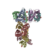
| ||||||||
|---|---|---|---|---|---|---|---|---|---|
| 1 | 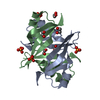
| ||||||||
| 2 | 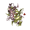
| ||||||||
| 3 | 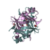
| ||||||||
| 4 | 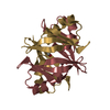
| ||||||||
| Unit cell |
|
-Components
| #1: Protein | Mass: 12244.663 Da / Num. of mol.: 8 Source method: isolated from a genetically manipulated source Source: (gene. exp.) Microcystis viridis (bacteria) / Production host: #2: Chemical | ChemComp-PO4 /   Mass: 94.971 Da / Num. of mol.: 6 / Source method: obtained synthetically / Formula: PO4 Mass: 94.971 Da / Num. of mol.: 6 / Source method: obtained synthetically / Formula: PO4#3: Chemical | ChemComp-EDO /   Mass: 62.068 Da / Num. of mol.: 13 / Source method: obtained synthetically / Formula: C2H6O2 Mass: 62.068 Da / Num. of mol.: 13 / Source method: obtained synthetically / Formula: C2H6O2#4: Water | ChemComp-HOH / |  Mass: 18.015 Da / Num. of mol.: 865 / Source method: isolated from a natural source / Formula: H2O Mass: 18.015 Da / Num. of mol.: 865 / Source method: isolated from a natural source / Formula: H2O |
|---|
-Experimental details
-Experiment
| Experiment | Method: X-RAY DIFFRACTION / Number of used crystals: 1 |
|---|
- Sample preparation
| Crystal | Density Matthews: 2.401 Å3/Da / Density % sol: 48.8 % |
|---|---|
| Crystal grow | Temperature: 295 K / Method: vapor diffusion, hanging drop / pH: 4 Details: Ammonium dihydrogen phosphate, pH 4.0, VAPOR DIFFUSION, HANGING DROP, temperature 295.0K |
-Data collection
| Diffraction | Mean temperature: 95 K |
|---|---|
| Diffraction source | Source: ROTATING ANODE / Type: RIGAKU RU200 / Wavelength: 1.5418 Å |
| Detector | Type: RIGAKU RAXIS IV / Detector: IMAGE PLATE / Date: Jan 15, 2005 |
| Radiation | Monochromator: GRAPHITE / Protocol: SINGLE WAVELENGTH / Monochromatic (M) / Laue (L): M / Scattering type: x-ray |
| Radiation wavelength | Wavelength: 1.5418 Å / Relative weight: 1 |
| Reflection | Resolution: 1.9→50 Å / Num. all: 73335 / Num. obs: 71428 / % possible obs: 97.4 % / Observed criterion σ(F): 2 / Observed criterion σ(I): 2 / Redundancy: 3.2 % / Biso Wilson estimate: 13.7 Å2 / Rmerge(I) obs: 0.092 |
| Reflection shell | Resolution: 1.9→1.97 Å / Redundancy: 3 % / Rmerge(I) obs: 0.347 / Mean I/σ(I) obs: 2.25 / Num. unique all: 6946 / % possible all: 95.3 |
- Processing
| Software |
| ||||||||||||||||||||||||||||||||||||
|---|---|---|---|---|---|---|---|---|---|---|---|---|---|---|---|---|---|---|---|---|---|---|---|---|---|---|---|---|---|---|---|---|---|---|---|---|---|
| Refinement | Method to determine structure: MIR / Resolution: 1.9→19.87 Å / Rfactor Rfree error: 0.003 / Data cutoff high absF: 460807.83 / Data cutoff low absF: 0 / Isotropic thermal model: RESTRAINED / Cross valid method: THROUGHOUT / σ(F): 0 / Stereochemistry target values: Engh & Huber
| ||||||||||||||||||||||||||||||||||||
| Solvent computation | Solvent model: FLAT MODEL / Bsol: 62.0686 Å2 / ksol: 0.368284 e/Å3 | ||||||||||||||||||||||||||||||||||||
| Displacement parameters | Biso mean: 20.2 Å2
| ||||||||||||||||||||||||||||||||||||
| Refine analyze |
| ||||||||||||||||||||||||||||||||||||
| Refinement step | Cycle: LAST / Resolution: 1.9→19.87 Å
| ||||||||||||||||||||||||||||||||||||
| Refine LS restraints |
| ||||||||||||||||||||||||||||||||||||
| LS refinement shell | Resolution: 1.9→2.02 Å / Rfactor Rfree error: 0.008 / Total num. of bins used: 6
| ||||||||||||||||||||||||||||||||||||
| Xplor file |
|
