 Movie
Movie Controller

[English] 日本語
 Yorodumi
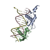
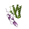
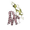
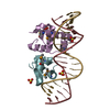
- PDB-1zg1: NarL complexed to nirB promoter non-palindromic tail-to-tail DNA site -
+ Open data

- Basic information
| Entry | Database: PDB / ID: 1zg1 | ||||||
|---|---|---|---|---|---|---|---|
| Title | NarL complexed to nirB promoter non-palindromic tail-to-tail DNA site | ||||||
 Components Components |
| ||||||
 Keywords Keywords | TRANSCRIPTION/DNA / protein-DNA complex / helix-turn-helix / two-component response regulator / dna bending / TRANSCRIPTION-DNA COMPLEX | ||||||
| Function / homology |  Function and homology information Function and homology informationDNA-binding transcription repressor activity / phosphorelay signal transduction system / nitrate assimilation / protein-DNA complex / transcription cis-regulatory region binding / DNA-binding transcription factor activity / regulation of DNA-templated transcription / DNA binding / ATP binding / identical protein binding / cytosol Similarity search - Function | ||||||
| Biological species |  | ||||||
| Method |  X-RAY DIFFRACTION / SYNCHROTRON / MOLECULAR REPLACEMENT / Resolution: 2.3 Å X-RAY DIFFRACTION / SYNCHROTRON / MOLECULAR REPLACEMENT / Resolution: 2.3 Å | ||||||
 Authors Authors | Maris, A.E. / Kaczor-Grzeskowiak, M. / Ma, Z. / Kopka, M.L. / Gunsalus, R.P. / Dickerson, R.E. | ||||||
 Citation Citation | Journal: Biochemistry / Year: 2005 Title: Primary and Secondary Modes of DNA Recognition by the NarL Two-Component Response Regulator. Authors: Maris, A.E. / Kaczor-Grzeskowiak, M. / Ma, Z. / Kopka, M.L. / Gunsalus, R.P. / Dickerson, R.E. #1: Journal: Nat.Struct.Biol. / Year: 2002Title: Dimerization allows DNA target site recognition by the NarL response regulator Authors: Maris, A.E. / Sawaya, M.R. / Kaczor-Grzeskowiak, M. / Jarvis, M.R. / Bearson, S.M.D. / Kopka, M.L. / Schroeder, I. / Gunsalus, R.P. / Dickerson, R.E. #2: Journal: Biochemistry / Year: 1996Title: Structure of the Escherichia coli response regulator NarL Authors: Baikalov, I. / Schroeder, I. / Kaczor-Grzeskowiak, M. / Grzeskowiak, K. / Gunsalus, R.P. / Dickerson, R.E. | ||||||
| History |
|
- Structure visualization
| Structure viewer | Molecule: MolmilJmol/JSmol |
|---|
- Downloads & links
-Download
| PDBx/mmCIF format | 1zg1.cif.gz | 115.1 KB | Display | PDBx/mmCIF format |
|---|---|---|---|---|
| PDB format | pdb1zg1.ent.gz | 85.3 KB | Display | PDB format |
| PDBx/mmJSON format | 1zg1.json.gz | Tree view | PDBx/mmJSON format | |
| Others |  Other downloads Other downloads |
-Validation report
| Arichive directory | https://data.pdbj.org/pub/pdb/validation_reports/zg/1zg1ftp://data.pdbj.org/pub/pdb/validation_reports/zg/1zg1 | HTTPS FTP |
|---|
-Related structure data
| Related structure data | 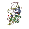 1zg5C  1je8S S: Starting model for refinement C: citing same article ( |
|---|---|
| Similar structure data |


-Links
 PDBj
PDBj


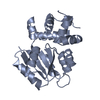
- Assembly
| Deposited unit | 
| ||||||||
|---|---|---|---|---|---|---|---|---|---|
| 1 | 
| ||||||||
| 2 | 
| ||||||||
| Unit cell |
| ||||||||
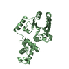
| Details | The biological unit is a dimer bound to DNA. There are two biological units in the asymmetric unit (chains A, B, C & D and chains E, F, G & H) |
-Components
| #1: DNA chain | Mass: 6124.965 Da / Num. of mol.: 2 / Source method: obtained synthetically / Details: solid phase synthesis #2: DNA chain | Mass: 6142.993 Da / Num. of mol.: 2 / Source method: obtained synthetically / Details: solid phase synthesis #3: Protein | Mass: 9827.909 Da / Num. of mol.: 4 / Fragment: DNA binding domain (residues 147-216) Source method: isolated from a genetically manipulated source Source: (gene. exp.) #4: Chemical | ChemComp-SO4 /   Mass: 96.063 Da / Num. of mol.: 9 / Source method: obtained synthetically / Formula: SO4 Mass: 96.063 Da / Num. of mol.: 9 / Source method: obtained synthetically / Formula: SO4#5: Water | ChemComp-HOH / |  Mass: 18.015 Da / Num. of mol.: 89 / Source method: isolated from a natural source / Formula: H2O Mass: 18.015 Da / Num. of mol.: 89 / Source method: isolated from a natural source / Formula: H2OHas protein modification | Y | |
|---|
-Experimental details
-Experiment
| Experiment | Method: X-RAY DIFFRACTION / Number of used crystals: 1 |
|---|
- Sample preparation
| Crystal | Density Matthews: 2.7 Å3/Da / Density % sol: 54.4 % | ||||||||||||||||||||
|---|---|---|---|---|---|---|---|---|---|---|---|---|---|---|---|---|---|---|---|---|---|
| Crystal grow | Temperature: 298 K / Method: vapor diffusion, hanging drop / pH: 7.5 Details: Ammonium sulfate, pH 7.5, hanging drop, temperature 298K, VAPOR DIFFUSION, HANGING DROP | ||||||||||||||||||||
| Components of the solutions |
|
-Data collection
| Diffraction | Mean temperature: 100 K |
|---|---|
| Diffraction source | Source: SYNCHROTRON / Site: NSLS  / Beamline: X8C / Wavelength: 1.1 Å / Beamline: X8C / Wavelength: 1.1 Å |
| Detector | Type: ADSC QUANTUM 4 / Detector: CCD / Date: Mar 11, 2001 |
| Radiation | Protocol: SINGLE WAVELENGTH / Scattering type: x-ray |
| Radiation wavelength | Wavelength: 1.1 Å / Relative weight: 1 |
| Reflection | Resolution: 2.25→25 Å / Num. all: 32539 / Num. obs: 32539 / % possible obs: 98.4 % / Observed criterion σ(I): -3 / Redundancy: 6.9 % / Biso Wilson estimate: 43.9 Å2 / Rmerge(I) obs: 0.088 / Rsym value: 0.067 / Χ2: 0.989 / Net I/σ(I): 18.3 |
| Reflection shell | Resolution: 2.25→2.3 Å / % possible obs: 98.8 % / Redundancy: 6.3 % / Rmerge(I) obs: 0.481 / Mean I/σ(I) obs: 3.9 / Num. measured obs: 1942 / Num. unique all: 1942 / Rsym value: 0.469 / Χ2: 1.047 / % possible all: 98.8 |
- Processing
| Software |
| |||||||||||||||||||||||||
|---|---|---|---|---|---|---|---|---|---|---|---|---|---|---|---|---|---|---|---|---|---|---|---|---|---|---|
| Refinement | Method to determine structure: MOLECULAR REPLACEMENT Starting model: PDB ENTRY 1JE8 Resolution: 2.3→9 Å / Cross valid method: THROUGHOUT / σ(F): 0 / Stereochemistry target values: Engh & Huber
| |||||||||||||||||||||||||
| Solvent computation | Bsol: 45.36 Å2 | |||||||||||||||||||||||||
| Displacement parameters | Biso mean: 45.475 Å2
| |||||||||||||||||||||||||
| Refine analyze |
| |||||||||||||||||||||||||
| Refinement step | Cycle: LAST / Resolution: 2.3→9 Å
| |||||||||||||||||||||||||
| Refine LS restraints |
| |||||||||||||||||||||||||
| LS refinement shell | Resolution: 2.3→2.44 Å / Rfactor Rfree error: 0.02
| |||||||||||||||||||||||||
| Xplor file |
|
