 Movie
Movie Controller


+ Open data

- Basic information
| Entry | Database: PDB / ID: 1zdl | ||||||
|---|---|---|---|---|---|---|---|
| Title | Crystal Structure of Mouse Thioredoxin Reductase Type 2 | ||||||
 Components Components | Thioredoxin reductase 2, mitochondrial | ||||||
 Keywords Keywords | OXIDOREDUCTASE / Selenocysteine / thioredoxin / reductase / flavoprotein | ||||||
| Function / homology |  Function and homology information Function and homology informationresponse to oxygen radical / Detoxification of Reactive Oxygen Species / thioredoxin-disulfide reductase (NADPH) / thioredoxin-disulfide reductase (NADPH) activity / hemopoiesis / cell redox homeostasis / flavin adenine dinucleotide binding / heart development / axon / neuronal cell body ...response to oxygen radical / Detoxification of Reactive Oxygen Species / thioredoxin-disulfide reductase (NADPH) / thioredoxin-disulfide reductase (NADPH) activity / hemopoiesis / cell redox homeostasis / flavin adenine dinucleotide binding / heart development / axon / neuronal cell body / dendrite / protein-containing complex binding / protein homodimerization activity / mitochondrion / cytoplasm / cytosol Similarity search - Function | ||||||
| Biological species |  | ||||||
| Method |  X-RAY DIFFRACTION / MOLECULAR REPLACEMENT / Resolution: 3 Å X-RAY DIFFRACTION / MOLECULAR REPLACEMENT / Resolution: 3 Å | ||||||
 Authors Authors | Biterova, E.I. / Turanov, A.A. / Gladyshev, V.N. / Barycki, J.J. | ||||||
 Citation Citation | Journal: Proc.Natl.Acad.Sci.Usa / Year: 2005 Title: Crystal structures of oxidized and reduced mitochondrial thioredoxin reductase provide molecular details of the reaction mechanism. Authors: Biterova, E.I. / Turanov, A.A. / Gladyshev, V.N. / Barycki, J.J. #1: Journal: Proc.Natl.Acad.Sci.USA / Year: 2001Title: Three-dimensional structure of a mammalian thioredoxin reductase: Implications for mechanism and evolution of a selenocysteine-dependent enzyme. Authors: Sandalova, T. / Zhong, L. / Lindqvist, Y. / Holmgren, A. / Schneider, G. #2: Journal: J.Mol.Biol. / Year: 1987Title: Refined structure of glutathione reductase at 1.54 A resolution. Authors: Karplus, P.A. / Schulz, G.E. | ||||||
| History |
|
- Structure visualization
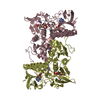
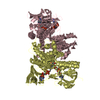
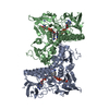
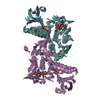
| Structure viewer | Molecule: MolmilJmol/JSmol |
|---|
- Downloads & links
-Download
| PDBx/mmCIF format | 1zdl.cif.gz | 111.9 KB | Display | PDBx/mmCIF format |
|---|---|---|---|---|
| PDB format | pdb1zdl.ent.gz | 83.7 KB | Display | PDB format |
| PDBx/mmJSON format | 1zdl.json.gz | Tree view | PDBx/mmJSON format | |
| Others |  Other downloads Other downloads |
-Validation report
| Arichive directory | https://data.pdbj.org/pub/pdb/validation_reports/zd/1zdlftp://data.pdbj.org/pub/pdb/validation_reports/zd/1zdl | HTTPS FTP |
|---|
-Related structure data
| Related structure data |  1zkqC  1h6vS S: Starting model for refinement C: citing same article ( |
|---|---|
| Similar structure data |


-Links
 PDBj
PDBj


- Assembly
| Deposited unit | 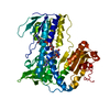
| ||||||||
|---|---|---|---|---|---|---|---|---|---|
| 1 | 
| ||||||||
| Unit cell |
|
-Components
| #1: Protein | Mass: 55816.562 Da / Num. of mol.: 1 Source method: isolated from a genetically manipulated source Source: (gene. exp.)  References: UniProt: Q9JLT4, thioredoxin-disulfide reductase (NADPH) |
|---|---|
| #2: Chemical | ChemComp-FAD /   Mass: 785.550 Da / Num. of mol.: 1 / Source method: obtained synthetically / Formula: C27H33N9O15P2 / Comment: FAD*YM Mass: 785.550 Da / Num. of mol.: 1 / Source method: obtained synthetically / Formula: C27H33N9O15P2 / Comment: FAD*YM |
| #3: Chemical | ChemComp-NDP /   Mass: 745.421 Da / Num. of mol.: 1 / Source method: obtained synthetically / Formula: C21H30N7O17P3 Mass: 745.421 Da / Num. of mol.: 1 / Source method: obtained synthetically / Formula: C21H30N7O17P3 |
| #4: Water | ChemComp-HOH /  Mass: 18.015 Da / Num. of mol.: 16 / Source method: isolated from a natural source / Formula: H2O Mass: 18.015 Da / Num. of mol.: 16 / Source method: isolated from a natural source / Formula: H2O |
| Has protein modification | Y |
-Experimental details
-Experiment
| Experiment | Method: X-RAY DIFFRACTION / Number of used crystals: 1 |
|---|
- Sample preparation
| Crystal | Density Matthews: 2.9 Å3/Da / Density % sol: 47 % |
|---|---|
| Crystal grow | Temperature: 291 K / Method: vapor diffusion, sitting drop / pH: 6.5 Details: 100 mM MES, 24% Peg 550 MME, pH 6.5, VAPOR DIFFUSION, SITTING DROP, temperature 291K |
-Data collection
| Diffraction | Mean temperature: 100 K |
|---|---|
| Diffraction source | Source: ROTATING ANODE / Type: RIGAKU / Wavelength: 1.5418 Å |
| Detector | Type: RIGAKU RAXIS IV / Detector: IMAGE PLATE / Date: Feb 26, 2005 |
| Radiation | Protocol: SINGLE WAVELENGTH / Monochromatic (M) / Laue (L): M / Scattering type: x-ray |
| Radiation wavelength | Wavelength: 1.5418 Å / Relative weight: 1 |
| Reflection | Resolution: 3→32.68 Å / Num. all: 12754 / Num. obs: 12754 / % possible obs: 100 % / Observed criterion σ(F): 0 / Observed criterion σ(I): 0 / Redundancy: 16.29 % / Rmerge(I) obs: 0.114 / Net I/σ(I): 17.8 |
| Reflection shell | Resolution: 3→3.11 Å / Redundancy: 16.66 % / Rmerge(I) obs: 0.431 / Mean I/σ(I) obs: 6.6 / % possible all: 100 |
- Processing
| Software |
| ||||||||||||||||||||||||||||||||||||||||||||||||||||||||||||||||||||||||||||||||
|---|---|---|---|---|---|---|---|---|---|---|---|---|---|---|---|---|---|---|---|---|---|---|---|---|---|---|---|---|---|---|---|---|---|---|---|---|---|---|---|---|---|---|---|---|---|---|---|---|---|---|---|---|---|---|---|---|---|---|---|---|---|---|---|---|---|---|---|---|---|---|---|---|---|---|---|---|---|---|---|---|---|
| Refinement | Method to determine structure: MOLECULAR REPLACEMENT Starting model: PDB entry 1H6V Resolution: 3→32.69 Å / Rfactor Rfree error: 0.009 / Data cutoff high absF: 182218.3 / Data cutoff low absF: 0 / Isotropic thermal model: RESTRAINED / Cross valid method: THROUGHOUT / σ(F): 0 / Stereochemistry target values: Engh & Huber
| ||||||||||||||||||||||||||||||||||||||||||||||||||||||||||||||||||||||||||||||||
| Solvent computation | Solvent model: FLAT MODEL / Bsol: 10.2389 Å2 / ksol: 0.289304 e/Å3 | ||||||||||||||||||||||||||||||||||||||||||||||||||||||||||||||||||||||||||||||||
| Displacement parameters | Biso mean: 61.9 Å2
| ||||||||||||||||||||||||||||||||||||||||||||||||||||||||||||||||||||||||||||||||
| Refine analyze |
| ||||||||||||||||||||||||||||||||||||||||||||||||||||||||||||||||||||||||||||||||
| Refinement step | Cycle: LAST / Resolution: 3→32.69 Å
| ||||||||||||||||||||||||||||||||||||||||||||||||||||||||||||||||||||||||||||||||
| Refine LS restraints |
| ||||||||||||||||||||||||||||||||||||||||||||||||||||||||||||||||||||||||||||||||
| LS refinement shell | Resolution: 3→3.19 Å / Rfactor Rfree error: 0.027 / Total num. of bins used: 6
| ||||||||||||||||||||||||||||||||||||||||||||||||||||||||||||||||||||||||||||||||
| Xplor file |
|
